Ethnicity Matters: Why Some Populations Face Heart Disease and Diabetes Earlier... Understanding the Biology Before Disease Appears
Ethnicity Matters: Why Some Populations Face Heart Disease and Diabetes Earlier
...Understanding the Biology Before Disease Appears
Summary
Before You Read This Article
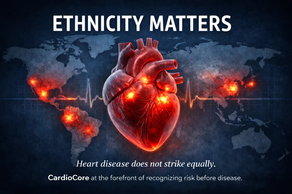
If you are South Asian, Indonesian, East or Southeast Asian, Hispanic (especially from Central and South America), Native American, African American, Caribbean, there is a very real chance that your risk for heart disease or diabetes is being underestimated.
Many people from these backgrounds are told they are healthy because they are not overweight, their cholesterol appears “normal,” their blood sugar is acceptable, or their stress test was reassuring. Yet family history often tells a different story—early heart attacks, diabetes, strokes, or sudden cardiovascular events in people who “seemed fine.”
This is not because these diseases strike randomly.
It is because they develop differently.
In many ethnic populations, cardiometabolic disease begins earlier, progresses faster, and does so at lower body weight. It is driven less by cholesterol alone and more by insulin resistance, inflammation, and subtle vascular injury that standard tests often miss.
As a result, risk goes unrecognized for years—until it announces itself “suddenly.”
There is almost never a “SUDDEN”… just no one was paying attention.
This article explains why ethnicity matters biologically, how metabolic stress quietly injures blood vessels long before symptoms appear, and why early recognition changes outcomes.
If you see yourself, or your family, in these words, that recognition matters.
Early awareness is not fear.
It is the point at which prevention becomes possible.
What follows is not about blame.
It is about seeing risk sooner so action can be taken before disease ever has a name.
Why I Wrote This
I wrote this article after seeing, and living through, too many stories of “sudden” heart attacks and premature diabetes in people who were repeatedly told they were fine. These events are rarely sudden. They reflect years of silent metabolic and vascular injury that can often be detected earlier—if we look with the right framework and the right tools.
Read On … This Is for You
Welcome. This was written for you. Please read on.
Hola — Esto es para ti. Sigue leyendo.
नमस्ते (Namaste) — यह आपके लिए है। कृपया आगे पढ़ें।
سلام (Salaam) — یہ آپ کے لیے ہے۔ براہِ کرم پڑھتے رہیں۔
你好 (Nǐ hǎo) — 这是为你而写的。请继续阅读。
안녕하세요 (Annyeonghaseyo) — 이 글은 당신을 위한 것입니다. 계속 읽어주세요.
Kamusta — Ito ay para sa iyo. Mangyaring magpatuloy sa pagbabasa.
Xin chào — Bài viết này dành cho bạn. Xin hãy đọc tiếp.
Bonjour — Ceci est pour vous. Continuez à lire.
Olá — Isto é para você. Continue lendo.
Welcome — This is for you. Please read on.
Risk Is Not About Blame — It’s About Biology, History, and Timing
Heart disease and diabetes are often labeled “lifestyle diseases,” implying that personal choice alone determines risk. While lifestyle matters, this explanation ignores substantial biological and epidemiologic evidence.³
Across the United States, African Americans and Caribbean populations, South Asians from India, Pakistan, and Bangladesh, Native Americans, Central and South American Hispanics, and East and Southeast Asian populations (including Chinese and Filipino individuals) develop cardiometabolic disease earlier in life and at lower body weight than individuals of European ancestry.⁴⁻⁷
This disparity persists even after accounting for access to care and socioeconomic factors.⁸
Clarifying the Role of Traditional Risk Factors
Recognizing metabolic and inflammatory drivers of disease does not negate the importance of traditional cardiovascular risk factors. Blood pressure, cholesterol, smoking status, physical activity, and family history remain essential components of risk assessment.
However, in many high-risk ethnic populations, these factors do not tell the full story when viewed in isolation. Normal cholesterol levels, acceptable blood pressure, or physical fitness can coexist with significant metabolic stress and early vascular disease.
True prevention requires focused attention, not only on traditional risk markers, but on how those markers behave within a broader metabolic and inflammatory context. Ethnicity provides critical insight into when and how aggressively risk should be evaluated.
“True medicine prioritizes wellness and prevention; the treatment of disease reflects a failure to prioritize those principles early.”
…CardioCore Metabolic Wellness Center
Built for Scarcity, Living in Abundance
Human metabolism evolved under conditions of intermittent food availability, high physical demand, and seasonal scarcity. The ability to efficiently store energy as fat was a profound survival advantage, allowing humans to endure famine, migration, illness, and environmental stress.⁹
For the vast majority of human evolution, periods of food excess were brief and infrequent—mere moments when viewed against the span of evolutionary time. Our biology was shaped almost entirely by scarcity, not abundance. Modern humans, however, still carry this ancient metabolic machinery, storing hundreds of thousands of excess calories, while now living in an environment defined by constant food availability, caloric density, and minimal physical demand.¹⁰
This mismatch between ancient biology and modern environment is not subtle. What once conferred survival advantage has become maladaptive. Cultural evolution has advanced at a pace far exceeding biological evolution, and our innate metabolic systems have not had time to recalibrate. The result is a predictable and accelerating rise in obesity, insulin resistance, and cardiometabolic disease.¹¹
Populations whose ancestors evolved with lower adipose storage capacity experience metabolic stress earlier when exposed to modern dietary patterns.¹² In regions where food sources were relatively stable and climates were warm and favorable, there was less evolutionary pressure to develop large subcutaneous fat reserves for insulation or prolonged energy storage.
As a result, energy storage capacity in these populations is often more limited, particularly in the subcutaneous compartment. After puberty and into early adulthood, the total number of fat cells in the body becomes relatively fixed for most individuals. When excess calories are consumed beyond immediate energy needs, that energy is therefore stored not by creating new fat cells (a process known as hyperplasia), but by enlarging existing fat cells, a process called hypertrophy.
In populations with reduced capacity for safe subcutaneous fat expansion, this limitation leads to earlier fat-cell hypertrophy, greater reliance on visceral fat storage, and a more rapid transition to metabolic dysfunction.
To understand why cardiometabolic disease appears earlier and at lower body weight in these populations, it is essential to examine not just how much fat is present, but where it is stored and how those fat cells respond under stress.
Not All Fat Is the Same: Subcutaneous vs Visceral Fat
Adipose tissue is a metabolically active endocrine organ, not inert storage.¹³
Subcutaneous fat, located beneath the skin, can expand through adipocyte hyperplasia—again, the creation of new fat cells—allowing relatively safe energy storage.¹⁴
Visceral fat, commonly known as “belly fat,” stored deep in the abdomen around vital organs, has limited capacity for hyperplasia and expands primarily through hypertrophy, the enlargement of existing cells.¹⁵ From an evolutionary perspective, this limitation is protective. Unlike subcutaneous fat, unrestricted expansion of visceral fat would compress vital organs and impair their function. As a result, biological constraints evolved to restrict fat-cell number in this compartment.
This protective design, however, comes at a cost in modern environments. When caloric excess persists, visceral fat cells are forced to enlarge beyond their optimal capacity, setting the stage for cellular stress, inflammation, and metabolic dysfunction.
Visceral fat accumulation is strongly associated with:
• Insulin resistance
• Inflammation
• Dyslipidemia
• Cardiovascular disease
• Premature mortality¹⁶⁻¹⁸
Importantly, individuals may have substantial visceral fat even at a “normal” body mass index (BMI).¹⁹ This mismatch between outward appearance and metabolic risk reflects patterns shaped by evolutionary genetics and helps explain why cardiometabolic disease appears earlier and at lower body weight in certain ethnic populations.
When Fat Cells Grow Too Large
Stress at the Cellular Level
As visceral fat cells enlarge, they not only outgrow their blood supply but experience marked mechanical stress on the cell membrane.²⁰
Basic physical principles apply: as the diameter of a structure increases, wall tension rises disproportionately. This is why large soap bubbles are unstable; they rupture.²¹
Similarly, hypertrophied adipocytes (fat cells) do not multiply indefinitely; instead, they enlarge, becoming progressively engorged and mechanically stressed. As their size increases, these cells develop:
Membrane strain — the fat cell enlarges beyond its normal capacity, placing increasing physical tension on the cell membrane and threatening its structural integrity
Reduced oxygen delivery (hypoxia) — expanding fat cells drift ever so slightly away from the microscopic blood vessels that normally nourish them, limiting oxygen and nutrient supply
Endoplasmic reticulum stress — meaning the cell’s internal protein-processing and quality-control system becomes overwhelmed, impairing normal cellular function and signaling that the cell is under metabolic distress
Activation of inflammatory signaling pathways — leading to the release of inflammatory cytokines, chemical messenger molecules that act as distress signals between cells, effectively “crying out for help” and alerting the immune system that the tissue is under threat²²⁻²⁴
These stressed adipocytes initiate adaptive survival responses designed to prevent cellular rupture and preserve tissue integrity. Chief among these responses is a reduction in insulin sensitivity, limiting further energy uptake into an already overfilled cell. While protective at the cellular level, this response has systemic consequences—propagating insulin resistance and inflammation throughout the body. As will be discussed later, the effects of this process are felt not only within other fat cells, but also in skeletal muscle and in specific regions of the brain that rely on insulin-mediated glucose uptake.
Why Fat Cells Stop Listening to Insulin
A common misconception is that insulin’s only job is to lower blood sugar. Insulin is produced by the pancreas in response to rising levels of glucose in the bloodstream. While insulin does help move sugar out of the blood, its far more important role is determining whether the body stores energy or burns it.
Insulin is the master regulator of metabolism.
When insulin levels are high, the body shifts into storage mode. Energy is stored as fat, fat burning is turned off, and metabolism slows. This happens most prominently after eating carbohydrates and when insulin remains elevated for long periods due to frequent eating or chronic stress.
When insulin levels are low, the body shifts into burning mode. Stored fat becomes available for energy, metabolism speeds up, and the body regains metabolic flexibility. In this state, the body is no longer storing energy—it is actively using it.
What raises insulin matters. Carbohydrates, especially refined carbohydrates and starches, raise insulin significantly. Eating frequently keeps insulin elevated for much of the day. In contrast, dietary fat alone has little effect on insulin levels and does not drive fat storage in the same way.
However, when carbohydrates are combined with fat, as is common in the Western diet but rarely encountered in nature, carbs win: insulin rises in response to the carbohydrates while the accompanying fat is efficiently stored. This combination creates a perfect metabolic storm—high insulin signaling storage, abundant incoming energy, suppressed fat burning, and accelerated fat accumulation. This distinction is critical.
Across many high-risk ethnic populations, including South Asian, East and Southeast Asian, Hispanic populations from Central and South America, and others, traditional diets are heavily centered around carbohydrates such as rice, potatoes, breads, and starches. When these dietary patterns are combined with modern food abundance and reduced physical activity, insulin remains chronically elevated, locking the body into storage mode and accelerating metabolic dysfunction.
Understanding insulin not as a “sugar hormone,” but as a fuel-allocation hormone, is essential to understanding why cardiometabolic disease develops earlier and more aggressively in certain populations.
The GLUT4 Door: How Insulin Resistance Spreads
Glucose enters many tissues through different transport systems. However, in adipose tissue, skeletal muscle, and certain key regions of the brain—including the hippocampus, which plays a central role in memory, learning, and cognitive function—glucose entry depends on insulin-mediated activation of GLUT4 (glucose transporter type 4).²⁷⁻²⁹
The term GLUT simply refers to glucose transport. There are multiple “doors” through which glucose can enter cells throughout the body, many of which do not require insulin. GLUT4 is unique. It is the only major glucose transporter that requires insulin to open the door, allowing glucose to move from the bloodstream into fat cells, muscle cells, and these insulin-sensitive regions of the brain.
For this reason, these tissues are disproportionately affected when insulin resistance develops. When insulin can no longer effectively activate GLUT4, glucose delivery to skeletal muscle and to insulin-sensitive brain regions becomes impaired—even when blood sugar levels appear normal. As will be discussed later, this helps explain why insulin resistance is increasingly recognized not only as a metabolic disorder, but also as a contributor to cognitive dysfunction, brain fog, and neurodegenerative conditions such as Alzheimer’s disease, sometimes described as a state of brain energy deprivation rather than a primary disorder of memory alone.
Skeletal muscle plays an especially critical role. As the body’s largest site of glucose disposal, muscle functions as the primary buffer for incoming carbohydrate—much like a central depository for excess fuel. When muscle becomes insulin resistant, even modest impairment markedly reduces the body’s ability to clear glucose from the bloodstream. Over time, this loss of metabolic “storage capacity” forces blood sugar levels higher and accelerates the progression from silent insulin resistance to overt metabolic disease.
Importantly, elevated blood glucose is a late manifestation of insulin resistance. Glucose itself is not the primary driver of early cardiometabolic dysfunction. That distinction belongs to insulin resistance and the chronic elevation of insulin that precedes it. While most individuals know their fasting glucose or hemoglobin A1c, insulin levels—the most sensitive predictor of future cardiometabolic disease—are rarely measured in routine clinical care.
Insulin acts as the molecular “key” that moves GLUT4 to the cell membrane, allowing glucose to enter the cell. Chronic hyperinsulinemia overwhelms this system, leading to:
• Impaired glucose uptake in skeletal muscle
• Reduced metabolic flexibility
• Compensatory insulin elevation
• Systemic insulin resistance³⁰
Because skeletal muscle is the largest site of glucose disposal in the body, insulin resistance at this level rapidly amplifies whole-body metabolic dysfunction.³¹
Insulin: The Switch Between Storing and Burning Energy
Insulin is the body’s primary regulator of energy allocation.³²
• High insulin promotes energy storage and suppresses fat oxidation
• Low insulin allows access to stored fat and metabolic flexibility³³
Dietary patterns strongly influence insulin dynamics. Carbohydrates, particularly refined carbohydrates, stimulate insulin secretion.³⁴ Frequent eating maintains insulin elevation, as insulin levels may remain elevated 6–8 hours after a meal.³⁵
With multiple daily meals and snacks, insulin rarely falls, locking the body into chronic storage mode.³⁶
The Hibernation Analogy: A System Gone Wrong
Seasonal hyperinsulinemia and fat accumulation are normal, adaptive processes in hibernating mammals such as bears, serving as preparation for prolonged periods of fasting.³⁷ In the autumn, bears deliberately consume diets rich in carbohydrates, particularly berries and honey, which raise insulin levels, promote rapid fat storage, and slow metabolic rate. This physiologic shift allows them to survive months of hibernation without food.
Humans, however, now live in a state of perpetual insulin elevation, without seasonal fasting or metabolic reset. Instead of a brief, adaptive pre-hibernation phase, modern dietary patterns—characterized by frequent eating and high carbohydrate availability—keep insulin chronically elevated throughout the year, all 365 days. This chronic state mirrors a continuous pre-hibernation physiology: fat storage is favored, fat burning is suppressed, and metabolism slows.³⁸
The result is not only weight gain, but widespread metabolic dysfunction. Importantly, in many individuals—particularly those with genetically limited capacity to expand fat-cell number—this dysfunction may occur with only modest or even minimal weight gain. In these cases, fat cells enlarge rather than multiply, accelerating cellular stress, insulin resistance, fatigue, and cardiometabolic disease despite the absence of overt obesity.
The Carb–Fat Trap: A Modern Metabolic Storm
Dietary fat alone has minimal effect on insulin secretion, whereas carbohydrates robustly stimulate insulin release.³⁹
This highlights a critical limitation of calorie-based thinking. Calories are a laboratory construct, measured outside the human body under controlled conditions. The body does not metabolize calories uniformly. It responds to fuel type, not calorie number. Carbohydrates, fats, and proteins are processed through distinct metabolic pathways, generate different hormonal responses, and produce markedly different effects on insulin signaling, fat storage, and energy expenditure.
This distinction has been demonstrated experimentally. When individuals consume diets matched for total calories but differing in macronutrient composition, those consuming lower-carbohydrate diets consistently exhibit higher daily energy expenditure—on the order of approximately 200–300 additional calories burned per day—compared with those consuming higher-carbohydrate diets.⁴⁰ This difference reflects hormonal regulation of metabolism rather than conscious effort or willpower.
In practical terms, the body does not simply “count calories.” It interprets incoming energy through hormonal signals—chief among them insulin—which determine whether energy is stored or burned. Thus, what we eat matters at least as much as how much we eat, particularly in populations already predisposed to insulin resistance and metabolic stress.
In practical terms:
• Fat consumption favors fat oxidation, allowing the body to burn stored energy and support a higher metabolic rate
• Carbohydrate consumption favors fat storage, particularly when intake is frequent or excessive, shifting the body toward energy conservation and a slower metabolic rate
The combination of high carbohydrate and high fat intake—common in Westernized diets—creates a uniquely harmful metabolic environment characterized by:
• Elevated insulin
• Suppressed fat oxidation
• Accelerated fat storage⁴⁰
Importantly, this combination is rarely encountered in nature. Traditional foods tend to be predominantly carbohydrate-based or fat-based, but not both in concentrated form. When carbohydrates raise insulin and fat is consumed simultaneously, insulin signals maximal energy storage while fat burning is effectively shut down. The result is a perfect metabolic storm: one that magnifies fat absorption, promotes rapid fat accumulation, and slows metabolism to its lowest functional state.
This combination is uncommon in natural diets but prevalent in modern processed foods.
In ethnic populations with traditionally high-carbohydrate diets, the addition of Western fats dramatically worsens metabolic risk.⁴¹⁻⁴³
What This Biology Is Telling Us
Insulin resistance is not a moral failing.
It is a biologically predictable response to:
• Cellular stress
• Chronic insulin exposure
• Evolutionary mismatch
In many ethnic populations, this process begins earlier, faster, and at lower body weight.
Understanding this biology is the foundation of prevention.
When Metabolic Stress Becomes Cardiovascular Disease
From Metabolic Stress to Vascular Injury
By the time insulin resistance and chronic inflammation are established, the body is no longer dealing with an isolated metabolic disturbance. What begins within stressed fat cells becomes a systemic process—one that injures blood vessels silently and progressively, often decades before diabetes is ever diagnosed.
This marks the true beginning of clinical disease: the long, quiet phase during which vascular injury accumulates, plaque biology becomes unstable, and risk steadily increases. Years later, this process may declare itself as a so-called “sudden” cardiac event—an outcome that appears unexpected only because the early warning signs were never recognized or acted upon.
The reality is not that these events occur without cause. They occur in plain sight. The biology is well described. The data are available. The technology to identify risk early already exists.
And yet far too often, modern medical practice does not apply established knowledge with the urgency and intensity that accepted standards require. Many physicians are not following established guidelines and are practicing as if time has stood still. Patients are reassured, under-imaged, under-evaluated metabolically, and frequently under-treated—despite decades of guideline-based standards that define what appropriate prevention and secondary prevention should look like.
System pressures may explain this environment—corporate medicine, time constraints, EHR demands—but they do not change the biological reality. When predictable disease becomes catastrophic, the tragedy is not only the event itself, but the lost opportunity to intervene when intervention could have changed the course entirely.
This is the fundamental distinction between reactive care and proactive medicine. Traditional models wait for disease to announce itself. Precision cardiometabolic medicine identifies risk long before symptoms appear and acts accordingly.
Heart disease does not begin with blocked arteries.
It begins with injured arteries.⁴⁴
The inner lining of blood vessels—the endothelium—is a living, responsive tissue. It regulates blood flow, blood pressure, clotting, immune signaling, and nutrient delivery. Predictably, chronic inflammation and hyperinsulinemia disrupt these functions early, silently, and progressively.⁴⁵ The outcome is not unexpected; it is simply too often unrecognized.
When insulin levels remain chronically elevated and inflammatory signals circulate continuously, normal vascular function begins to break down in predictable ways. One of the earliest changes is a reduction in nitric oxide availability. Nitric oxide is a critical molecule produced by the lining of blood vessels that allows arteries to relax, maintain healthy blood flow, and resist inflammation. Under normal conditions, insulin helps stimulate nitric oxide production, supporting vascular health.
In states of insulin resistance and chronic inflammation, this protective pathway is impaired. Blood vessels lose their ability to dilate appropriately, oxidative stress increases, and the vascular environment becomes increasingly hostile. At the same time, inflammatory signaling promotes the recruitment of immune cells into the vessel wall, where they contribute to endothelial injury, plaque formation, and plaque instability.⁴⁶
This combination—reduced nitric oxide, increased oxidative stress, and immune activation—creates the conditions for atherosclerosis to develop silently and progress long before symptoms or abnormal routine tests appear.
This environment is fertile ground for atherosclerosis, even when cholesterol levels appear “acceptable.” In certain ethnic populations, underlying metabolic and inflammatory susceptibility makes this ground especially fertile.
Why Cholesterol Alone Fails to Explain Risk
For decades, cardiovascular risk has been framed almost entirely through cholesterol levels. While cholesterol plays a role, this narrow focus has created a dangerous blind spot—particularly in high-risk ethnic populations, where cardiometabolic disease often develops in the absence of markedly elevated LDL cholesterol.
Insulin resistance profoundly alters how cholesterol behaves, not merely how much of it circulates. Under normal physiological conditions, cholesterol is not harmful. It is an essential molecule, integral to cell membrane structure, a precursor for all steroid hormones (including cortisol, aldosterone, estrogen, and testosterone), and a key component of mitochondrial energy pathways through compounds such as ubiquinone (coenzyme Q10). Cholesterol also plays important roles in immune function and, in healthy metabolic states, may exert anti-inflammatory effects.
The problem arises when the metabolic environment changes.
In states of chronic hyperinsulinemia and inflammation, cholesterol is transformed from a structural and functional molecule into a participant in vascular injury. Insulin resistance disrupts hepatic lipid handling, alters lipoprotein metabolism, and changes the inflammatory environment within blood vessels. Cholesterol particles circulate longer, interact with inflamed endothelium, and become incorporated into an already hostile vascular milieu.⁴⁷⁻⁴⁹
Critically, in many high-risk ethnic populations, atherosclerosis is not primarily an LDL-driven disease. Instead, LDL becomes involved downstream—recruited into a vascular environment already shaped by insulin resistance, endothelial dysfunction, and chronic immune activation. Within this metabolically toxic milieu, LDL particles are more likely to become smaller, denser, and susceptible to oxidative modification—not because LDL is inherently harmful, but because the surrounding biology has already shifted toward inflammation and instability.
This distinction explains a devastating and commonly misunderstood clinical pattern seen repeatedly in high-risk ethnic populations: individuals with “normal” or acceptable LDL cholesterol, no flow-limiting obstruction on stress testing, no warning symptoms—and yet a myocardial infarction. In these cases, the underlying disease was never excess cholesterol alone. It was inflammation and insulin resistance driving unstable plaque biology long before cholesterol values appeared abnormal.⁵⁰
The Consequence of a Cholesterol-Centric Bias
Because LDL cholesterol is often not markedly elevated at the time coronary disease is discovered in these patients, many are inappropriately treated with low-intensity statin therapy, despite clear evidence and long-standing guideline recommendations to the contrary.
No major cardiovascular society has ever endorsed low-dose statin therapy for secondary prevention—defined by the presence of established atherosclerotic disease—irrespective of baseline or post-treatment cholesterol levels.
Since the 2013 ACC/AHA cholesterol guidelines, secondary prevention has been based on statin intensity and percent LDL reduction, not absolute LDL values. This shift reflects an important biological reality: in established atherosclerotic disease, the benefit of statin therapy is related to its pleiotropic, anti-inflammatory, and plaque-stabilizing effects—not merely its ability to lower cholesterol numbers.
Further underscoring this point, diabetes has been classified as a coronary heart disease risk equivalent, meaning that an individual with diabetes carries a cardiovascular risk comparable to someone who has already suffered a myocardial infarction. As such, current guidelines recommend high-intensity statin therapy in patients with diabetes, irrespective of baseline LDL cholesterol. Yet, in clinical practice, this standard is frequently ignored or inadequately applied.
The consequence is not trivial. Treating a patient with established or equivalent cardiovascular risk using subtherapeutic statin dosing is analogous to selling a car without an airbag, despite knowing that safety standards require one. Established standards of care exist precisely to prevent catastrophic outcomes. When those standards are not followed, patients are exposed to avoidable risk without their informed consent.
Physicians carry a responsibility not only to prescribe, but to inform. If evidence-based therapy is being withheld or modified, patients deserve to know why. Too often, however, individuals are reassured, under-treated, and left unaware that their care falls short of accepted medical standards. This is not personalized medicine—it is missed medicine.
And when disease inevitably progresses, it is the patient who pays the price for a failure of diligence that could, and should, have been prevented.
The therapeutic benefit in this setting has little to do with treating cholesterol itself and everything to do with addressing the inflammatory and plaque-stabilizing effects associated with adequate statin intensity. Treating established coronary disease with low-dose statins reflects a fundamental misunderstanding of the disease process and represents another missed opportunity to mitigate risk.
More importantly, an exclusive focus on cholesterol distracts clinicians from the true drivers of atherosclerosis: chronic inflammation, insulin resistance, and metabolic dysfunction. This bias has measurable consequences. In acute coronary syndromes, approximately 70% of patients presenting to emergency departments have LDL cholesterol levels that fall within “normal” or “acceptable” ranges. Yet the narrative persists that cholesterol was the primary problem.
Once again, the disease was present. The biology was active. The warning signs existed.
What was missing was attention.
When cholesterol is blamed in isolation, the real pathology remains unaddressed, and the window for meaningful prevention closes.
The Myth of the “Big Blockage”
One of the most counterintuitive truths in cardiology is this:
Nearly two-thirds of heart attacks occur in arteries narrowed less than 50%.⁵¹
These are not large, fixed blockages. They are inflamed, unstable plaques that rupture suddenly.
Stress tests are designed to detect flow-limiting obstructions—not inflammatory plaque. As a result, many high-risk individuals are falsely reassured, particularly when they are younger, thinner, or have normal cholesterol.⁵²
This limitation disproportionately harms ethnic populations whose disease is driven more by metabolic inflammation than by long-standing obstructive disease.
Why Heart Disease Often Appears Before Diabetes
Many people assume diabetes must come first and heart disease follows later. In reality, the cardiovascular system often suffers damage years before diabetes is diagnosed.⁵³
Long before blood sugar rises, blood vessels are subjected to a relentless metabolic assault. They are bathed continuously in elevated insulin levels, inflammatory signaling molecules, circulating free fatty acids, and oxidative stress. All of this occurs quietly, often while blood glucose remains “normal,” sustained only by excessive insulin production. This is precisely why hemoglobin A1C, a late and blunt marker, so often fails to detect early disease—allowing vascular injury to progress unnoticed for years.⁵⁴
By the time diabetes is diagnosed, vascular injury is frequently advanced. In high-risk ethnic populations, this silent phase begins earlier and progresses faster.
Why These Patterns Are Especially Dangerous in Certain Ethnic Groups
Certain ethnic populations are biologically predisposed to develop cardiometabolic disease earlier in life due to a convergence of factors, including lower visceral fat storage capacity, earlier fat-cell hypertrophy, higher habitual carbohydrate intake, and more pronounced insulin responses to dietary carbohydrate. Together, these characteristics result in a longer lifetime exposure to insulin resistance and chronic inflammation. Over time, this extended metabolic stress accelerates plaque formation, impairs endothelial function, and advances vascular injury—leading to cardiovascular events that often occur 10 to 20 years earlier than expected, even in the absence of traditional warning signs.⁵⁵⁻
This is not because these individuals are less healthy. It is because their biology reaches its limits sooner in a modern environment.
Seeing Under the Hood: Why Early Detection Matters
If disease begins silently, waiting for symptoms is the wrong strategy. No one should suffer a heart attack without ever knowing what their heart looked like beforehand. For individuals from higher-risk ethnic backgrounds, earlier evaluation is not aggressive—it is appropriate.
One of the most valuable tools for early detection is coronary artery calcium (CAC) scoring, which identifies calcified plaque as a marker of cumulative arterial injury and chronic inflammation.⁵⁸ A zero score is reassuring, but it is not permanent. A non-zero score is not a sentence—it is a signal. It tells us that atherosclerosis has already begun and that the metabolic drivers of disease require attention.
The most common failure in clinical practice is an act of omission: failing to obtain direct coronary imaging, such as a calcium score or coronary CT angiography, when risk is present. Equally harmful is an act of commission: ordering a stress test and using a “normal” result to falsely reassure patients that their heart is healthy.
Stress testing does not evaluate whether coronary disease is present. It evaluates whether blood flow is severely impaired under stress—that is, whether a large, fixed obstruction is limiting oxygen delivery. In practical terms, stress testing detects only advanced disease, typically when arterial narrowing exceeds 70 percent. Yet the majority of heart attacks do not arise from these severe blockages. They occur when smaller, inflamed, unstable plaques rupture—lesions that stress testing is not designed to detect.
By contrast, coronary calcium scoring answers fundamentally different and far more relevant questions:
Is atherosclerosis present?
How extensive is it?
How urgent is intervention?
Increasingly, this same inflammatory and insulin-resistant vascular environment has been linked not only to cardiovascular disease, but also to heightened risk for cognitive decline and dementia, chronic kidney disease, fatty liver disease, and even certain cancers—underscoring that atherosclerosis is not an isolated cardiac condition but a manifestation of systemic metabolic dysfunction.
Stress testing cannot provide this information. It does not assess plaque burden, plaque biology, or systemic disease activity. When stress testing is used as a screening tool rather than a tool for evaluating symptoms, it too often delays recognition of disease until prevention is no longer possible.
This pattern—failing to image when risk is present, and falsely reassuring when stress tests are normal—represents a recurring combination of omission and commission. The result is predictable, preventable disease that continues to progress quietly until it declares itself as an “unexpected” event.
Why Metabolic Testing Must Follow Imaging
Once early plaque is identified, the most important question is no longer “How blocked are the arteries?” but rather, “Why is disease already developing?” This is where metabolic testing becomes essential.
A two-hour oral glucose tolerance test (OGTT) with fasting, one-hour, and two-hour insulin measurements is a simple, widely available test that can uncover insulin resistance years before diabetes is diagnosed—even when fasting glucose and hemoglobin A1C remain normal.⁵⁹⁻⁶¹ This same test is routinely used in pregnancy to detect gestational diabetes, underscoring both its safety and clinical utility. Yet outside of obstetrics, it is rarely employed—particularly in cardiology—despite substantial evidence demonstrating its value in the cardiology literature, in patients with atherosclerosis and “normal” blood sugar.
When performed correctly, this test reveals critical metabolic information, including hyperinsulinemia, impaired insulin clearance, and early metabolic dysfunction—abnormalities that often precede overt disease by many years. Without this insight, cardiometabolic risk remains hidden, and opportunities for prevention are missed.
Another underutilized marker is the triglyceride-to-HDL cholesterol ratio. A ratio above approximately 2.5 strongly suggests underlying insulin resistance, regardless of LDL cholesterol levels.⁶² Importantly, this information is not hidden or specialized—it is readily available on the standard lipid panel routinely ordered in clinical practice. Its value, however, depends entirely on proper interpretation. When viewed through a purely cholesterol-centric lens, this signal is often overlooked, despite its ability to reveal early metabolic risk long before traditional markers become abnormal. Its utility is limited not by availability, but by whether clinicians recognize what it signifies.
From a preventive standpoint, early coronary imaging provides the necessary context for interpreting these metabolic findings. In individuals from high-risk ethnic populations, particularly those over the age of 40, or younger in the presence of strong family history, coronary artery calcium (CAC) scoring offers a low-risk, high-yield method of identifying early atherosclerosis. The test is relatively inexpensive, widely available, and carries radiation exposure comparable to, or only slightly greater than, that of a screening mammogram.
When combined, coronary calcium scoring and a two-hour OGTT with insulin response form a powerful foundation for early risk detection. If both evaluations are normal, reassurance is appropriate, and repeat testing can be considered in five years, depending on overall risk. If either is abnormal, it signals an opportunity—often years before symptoms—to intervene proactively and alter the disease trajectory.
Clinical judgment remains essential. In women from high-risk ethnic populations, a baseline coronary calcium assessment around age 50 is reasonable and appropriate, reflecting the partial cardioprotective effects of endogenous estrogen prior to menopause. However, this protection is not absolute and does not eliminate risk—particularly in the presence of strong family history, insulin resistance, diabetes, or other markers of metabolic dysfunction. In such cases, earlier evaluation—often between ages 40 and 45—is both justified and prudent, as metabolic risk may overwhelm hormonal protection long before menopause.
In an ideal system, this approach would represent standard care. Instead, despite mounting evidence, medicine too often remains reactive—waiting for disease to declare itself rather than identifying risk when intervention is most effective. The tools are available. The data are compelling. What remains is the willingness to apply them thoughtfully and early, before preventable disease becomes irreversible.
From Late Intervention to Early Understanding
The purpose of early evaluation is not fear—it is clarity. Identifying risk before disease becomes clinically apparent allows for thoughtful, proportional action rather than crisis-driven decisions made after damage has already occurred. This distinction is especially critical in ethnic populations in whom cardiometabolic disease tends to develop earlier, progress more quietly, and remain underestimated by traditional screening approaches. When risk is recognized at its biological origins, outcomes can change—often without medications, procedures, or emergency interventions.
True prevention requires translating biological insight into meaningful action. Rather than waiting for disease to announce itself, the focus must shift to understanding why risk is present and how it is evolving—while intervention remains effective. This requires a precision, personalized approach—one that moves beyond population averages to account for individual biology. Such an approach integrates genetic information to clarify inherited susceptibility; advanced metabolic evaluation to assess insulin dynamics, energy production, and mitochondrial stress; and hormonal assessment to understand how endocrine balance shapes the cardiometabolic terrain. It also incorporates comprehensive gut microbiome analysis to identify inflammatory and metabolic disruption, along with nutrition guided by biological response rather than generic trends.
Equally important are the non-biochemical drivers of metabolic health—stress physiology, sleep quality, social connection, and a sense of meaning—because metabolism does not exist in isolation from life itself.
This work is not about chasing laboratory numbers or suppressing symptoms after the fact. It is about addressing root causes early, while there is still time to restore balance—metabolically, hormonally, and systemically—and to change the trajectory of disease before it becomes irreversible.
Timing Is the Difference Between Prevention and Regret... Risk Is Invisible—Until It Isn’t
Ethnicity matters… but not in isolation.
Certain populations carry well-documented cardiometabolic vulnerability, and ignoring those biological realities costs lives. But the deeper truth is this: genetic risk is not confined to ethnic labels. Because of genetic diversity, migration, and shared ancestral traits, many individuals who do not belong to traditionally high-risk groups carry the same internal metabolic patterns—insulin resistance, visceral adiposity, inflammation, impaired cellular energy, and early vascular injury—despite looking entirely different on the outside.
You may not “look” high risk.
You may not fit the stereotype.
Yet on the inside, your biology may follow the same trajectory.
This is why family history matters—but not as a sentence. It is a signal. One that reflects inherited susceptibility interacting with lived culture, environment, and time. Genetics load the framework, but they do not dictate the outcome. How risk is expressed is shaped by nutrition, physical activity, stress physiology, sleep quality, social connection, and meaning—factors that quietly influence biology long before disease has a name.
When heart disease or diabetes appears “suddenly,” it is almost never sudden. It represents years—often decades—of silent metabolic and vascular injury that standard screening fails to detect. Normal cholesterol, normal blood sugar, and reassuring stress tests do not guarantee protection, particularly in individuals whose disease develops earlier, faster, and below conventional thresholds.
True prevention is quiet.
It happens before symptoms.
Before procedures.
Before emergencies.
This entire article is guided by one essential principle:
Risk must be recognized early enough to change its course.
This is precisely where precision cardiometabolic medicine becomes essential.
At CardioCore Metabolic Wellness Center, we do not guess, and we do not rely on population averages. We evaluate the full biological terrain using advanced, clinically grounded tools, including:
• Genetic testing to identify inherited susceptibility and guide personalized risk mitigation strategies
• Advanced metabolic testing to assess insulin dynamics, glucose handling, fuel utilization, nutrient status, and mitochondrial function
• Hormonal evaluation to understand endocrine balance and its downstream effects on cardiometabolic regulation
• Comprehensive gut microbiome analysis to identify inflammatory, immune, and metabolic drivers of disease
• Detailed assessment of lipid physiology, inflammation, and vascular health beyond standard cholesterol panels
• Individualized nutritional strategies informed by biological response rather than one-size-fits-all recommendations
• Evaluation of stress physiology, sleep quality, social connection, and meaning, recognizing that metabolism does not exist in isolation from life itself
These data are not collected for curiosity. They are used to design precise, personalized strategies aimed at restoring metabolic balance, improving cellular energy, and reducing long-term cardiometabolic risk before irreversible disease takes hold.
This approach allows us to address the full complexity of cardiometabolic disease and its systemic consequences—including diabetes, heart disease, lipid disorders, weight dysregulation, chronic inflammation, and impaired energy production. We also recognize that these same metabolic disturbances underlie many conditions traditionally viewed as separate, including neurodegenerative disease and certain cancers.
This is not about managing disease once damage is done.
It is not about reacting after biology has already declared itself.
It is about timing.
If you recognized yourself in this article—your family history, your culture, or your own health trajectory—do not dismiss that recognition. Biology is signaling for attention.
This level of precision, integration, and personalization does not exist in conventional care. And for individuals at elevated risk, it is no longer optional.
Because prevention is not passive.
And when it comes to cardiometabolic disease, waiting costs lives.
If you may be at risk, the time to act is now.
The Time to Act is Now
Hola — El momento de actuar es ahora.
नमस्ते (Namaste) — अब कार्य करने का समय है।
سلام (Salaam) — اب عمل کرنے کا وقت ہے۔
你好 (Nǐ hǎo) — 现在是采取行动的时候。
안녕하세요 (Annyeonghaseyo) — 지금이 행동할 때입니다.
Kamusta — Ngayon na ang oras para kumilos.
Xin chào — Đã đến lúc hành động.
Bonjour — Le moment d’agir, c’est maintenant.
Olá — O momento de agir é agora.
Welcome — The time to act is now.
A Note to the Reader
This article is written in dedication to Harry who inspired me to write this, and to his wife’s uncle, who immigrated to the United States from India and passed away “suddenly” from a heart attack at the age of 55.
It is also dedicated to Vinod, whose life was lost prematurely due to the rupture of a diseased artery—an event that was described as acute, but which almost certainly reflected years of silent metabolic and vascular injury.
At the same time, this article is dedicated to Vinay, also from India, who, through earlier recognition and intervention, is doing well—serving as a powerful reminder that outcomes can change when risk is identified in time.
Finally, this article is written in memory of Bradley’s father Blas, from the Philippines, who suffered early cardiovascular death despite appearing physically fit and otherwise healthy.
These stories are not isolated tragedies. They represent missed opportunities for earlier recognition, deeper understanding, and more precise prevention.
Heart disease is often described as sudden. Medically and biologically, it rarely is. It develops quietly over years—often decades—before the event occurs.¹
This article is not written to assign blame.
It is written to promote recognition.
Each of these lives represents an opportunity—not only to honor those lost, but to learn, refine our approach, and move forward with greater clarity. Prevention begins with seeing risk early, understanding it fully, and acting with intention.
Author: Dr John Sciales
Director, CardioCore Metabolic Wellness Center
"Getting to the Core- where being Healthy is Not an Accident"
Click here to book a discovery call
Click here to join our private community
Click here to speak with our CardioMetabolic virtual assistant
References
Libby P. Inflammation in atherosclerosis. Nature. 2002.
Yusuf S et al. Global burden of cardiovascular disease. Circulation. 2020.
Mozaffarian D et al. Lifestyle and cardiometabolic risk. NEJM. 2016.
Misra A, Ganda OP. South Asians and insulin resistance. J Clin Endocrinol Metab. 2007.
Enas EA et al. Coronary artery disease in Asian Indians. Indian Heart J. 2019.
Haffner SM et al. Ethnic differences in insulin resistance. Diabetes Care. 1996.
Narayan KMV et al. Native American diabetes epidemic. JAMA. 1998.
Diez Roux AV. Race, socioeconomic position, and CVD. Circulation. 2012.
Neel JV. Thrifty genotype hypothesis. Am J Hum Genet. 1962.
Speakman JR. Evolutionary perspectives on obesity. Obes Rev. 2008.
Wells JCK. Mismatch theory and metabolic disease. Int J Epidemiol. 2012.
Yajnik CS. Thin–fat Indian phenotype. Int J Obes. 2003.
Kershaw EE, Flier JS. Adipose tissue as endocrine organ. J Clin Endocrinol Metab. 2004.
Spalding KL et al. Adipocyte turnover in humans. Nature. 2008.
Tchkonia T et al. Mechanisms of adipose hypertrophy. Endocr Rev. 2010.
Després JP. Visceral obesity and cardiometabolic risk. Circulation. 2012.
Fox CS et al. Visceral adiposity and CVD. Circulation. 2007.
Kuk JL et al. Visceral fat and mortality. Obesity. 2006.
Shah RV et al. Normal BMI, high visceral fat. J Am Coll Cardiol. 2014.
Sun K et al. Adipose tissue hypoxia. J Clin Invest. 2011.
Laplace P-S. Law of surface tension. 1805.
Ye J. Adipose tissue inflammation. J Clin Invest. 2009.
Hotamisligil GS. ER stress and metabolic disease. Cell. 2010.
Cinti S et al. Adipocyte death and inflammation. Obesity. 2005.
Smith U. Insulin resistance in fat cells. Diabetologia. 2002.
Shulman GI. Insulin resistance mechanisms. J Clin Invest. 2000.
Bryant NJ et al. GLUT4 trafficking. Nat Rev Mol Cell Biol. 2002.
McEwen BS. Metabolic effects in hippocampus. Nat Neurosci. 2007.
Arnold SE et al. Brain insulin resistance. Diabetes. 2018.
DeFronzo RA. Pathogenesis of insulin resistance. Diabetes. 2009.
Richter EA, Hargreaves M. Muscle glucose uptake. Physiol Rev. 2013.
Saltiel AR, Kahn CR. Insulin signaling. Nature. 2001.
Cahill GF. Fuel metabolism. N Engl J Med. 1970.
Ludwig DS. Glycemic index and insulin. JAMA. 2002.
Boden G et al. Prolonged insulin elevation. Am J Physiol. 1996.
Mattson MP et al. Meal timing and metabolism. NEJM. 2014.
Donahue SW et al. Hibernation metabolism. Physiology. 2006.
Lustig RH. Insulin resistance and chronic disease. Prog Cardiovasc Dis. 2014.
Hall KD et al. Macronutrients and insulin. Cell Metab. 2018.
Taubes G. Carbohydrate–insulin model. BMJ. 2021.
Willett WC et al. Dietary patterns and CVD. Lancet. 2019.
Hu FB. Refined carbohydrates and metabolic disease. AJCN. 2010.
Enas EA et al. Dietary transitions in South Asians. Curr Diab Rep. 2014.
Ross R. Atherosclerosis—an inflammatory disease. N Engl J Med. 1999.
Libby P. Mechanisms of acute coronary syndromes. Nature. 2013.
Vanhoutte PM et al. Endothelial dysfunction. Eur Heart J. 2017.
Reaven GM. Insulin resistance and cardiovascular disease. Diabetes Care. 2011.
Krauss RM. Atherogenic lipoprotein phenotype. Circulation. 2004.
Borén J et al. LDL retention and inflammation. J Intern Med. 2020.
Ridker PM. Inflammation, CRP, and vascular disease. Circulation. 2016.
Falk E et al. Plaque rupture mechanisms. Circulation. 1995.
Shaw LJ et al. Limitations of stress testing. J Am Coll Cardiol. 2009.
Haffner SM et al. Cardiovascular risk before diabetes. N Engl J Med. 1998.
Tabák AG et al. Prediabetes and vascular risk. Lancet. 2012.
Yudkin JS et al. Ethnicity and insulin resistance. Diabetologia. 1999.
Lear SA et al. Ethnic differences in visceral adiposity. Circulation. 2012.
Tillin T et al. South Asians and early CVD. J Am Coll Cardiol. 2013.
Budoff MJ et al. Coronary calcium and risk prediction. J Am Coll Cardiol. 2018.
Kraft JR. Hyperinsulinemia and heart disease. J Insulin Resist. 1975.
Dankner R et al. OGTT insulin patterns. Diabetes Care. 2009.
Reaven GM. Insulin resistance syndrome. Annu Rev Med. 2003.
McLaughlin T et al. TG/HDL ratio and insulin resistance. Am J Cardiol. 2005.
