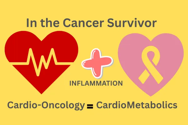
Cardio-Oncology and the Cardiometabolic Responsibilities in the Cancer Survivor
Cardio-Oncology and the Cardiometabolic Responsibilities in the Cancer Survivor
Dr. John Sciales
Founder & Director, CardioCore Metabolic Wellness Center
This article was written for a medical journal in response to what I, as a cardiometabolic and functional medicine physician, have witnessed happening in cardio-oncology. When you see something, you must say something. I cannot stand by while critical metabolic and inflammatory risks in cancer survivors are ignored. As a cardiometabolic expert and functional physician, I am writing this to challenge the status quo, to make care better for our population — and to save lives. Each of us needs to step forward and speak up to save lives and make a difference.
Due to the length and depth of this article, I have divided it into 13 sections. Each section addresses a critical dimension of cardio-oncology and cardiometabolic health, including issues from a functional medicine point of view. Depending on your level of expertise or area of interest, you may choose to focus on the sections most relevant to your clinical practice, research, or integrative approach to care.
Acknowledgment:
I would like to express my deep gratitude to Dr. Paul Ridker for his pioneering research identifying inflammation as a central driver of cardiovascular disease. I had the privilege of meeting Dr. Ridker at the national Cardiometabolic Health Congress, and beyond being a visionary scientist and humanitarian, he is a true gentleman; an individual who practices what he preaches. He embodies the principles he teaches, not only through his work but also through his own personal health and athletic lifestyle. His life and leadership are a living testament to the principles of prevention and resilience, setting an example for all of us in medicine to follow. https://prevmed.bwh.harvard.edu/paul-m-ridker-md-mph/
The author also gratefully acknowledges the contributions of Dr. Christie M. Ballantyne, M.D., Professor of Medicine and Chief of the Section of Cardiovascular Research at Baylor College of Medicine, a world recognized authority in preventive cardiology, lipid metabolism, and cardiovascular risk reduction. His expertise in in atherosclerosis imaging, inflammatory biomarkers, and cardiometabolic prevention have been instrumental in advancing the understanding of terrain based cardiovascular risk and precision prevention in oncology and cardiometabolic medicine. As a teacher, mentor, physician and humanitarian, he has been paving the way to recognize and reduce the CardioMetabolic burden; saving lives through science, compassionate and inspiration. https://www.bcm.edu/people-search/christie-ballantyne-17846
Abstract
Background:
Cancer survivors now face a new and often unrecognized threat: the insidious rise of insulin resistance, inflammation, and cardiometabolic dysfunction triggered by chemotherapy, immunotherapy, radiation, and the terrain-altering effects of survivorship itself.
Objective:
To establish that the cardio-oncologist must assume full responsibility for the metabolic, inflammatory, and cardiovascular risks in the post-cancer patient — risks that are not secondary, but central to survivorship outcomes.
Key Findings:
Chemotherapy and immunotherapy induce a pro-inflammatory, insulin-resistant state, increasing the incidence of new-onset diabetes and accelerating atherosclerosis and heart failure. Many cancer survivors consume excess calories to “feel safe,” unknowingly fueling hyperinsulinemia and metabolic dysfunction.
GLUT1 and GLUT3 glucose transporters, which are insulin-independent, allow cancer cells to thrive in hyperglycemic environments — and high glucose states driven by insulin resistance support both cancer recurrence and cardiovascular decline. Standard labs and stress tests miss early terrain dysfunction.
Precision cardiometabolic testing — including 2-hour OGTT with insulin, CRP-hs, CAC scoring, FFRct imaging, and micronutrient panels — is essential. Cardiologists who ignore insulin resistance, mitochondrial health, and metabolic terrain are treating disease with one hand tied behind their back.
Conclusion:
The role of the cardio-oncologist is no longer limited to monitoring for ischemia, LVEF or managing arrhythmia. It is about restoring vitality, preserving life, and extinguishing the inflammatory fire that silently fuels both cardiovascular disease and cancer recurrence. This is not someone else’s job. It is the core responsibility of the cardio-oncologist — the new conductor of survivorship care.
I have separated this article into 13 sections with the references after each section.
Section 1. Introduction
Cancer is now survivable, but the aftermath of treatment often leaves patients with invisible wounds: cardiometabolic dysfunction, inflammation, and mitochondrial injury. Cardiologists must look beyond cholesterol and coronary anatomy. They must understand how cancer therapies disrupt metabolic signaling, damage vascular integrity, and shift cardiac energy utilization from glucose and ketones to toxic lipid intermediates.
Section 2. The Post-Cancer Survivor’s Cardiometabolic Burden
Chemotherapy and immunotherapy create a state of systemic inflammation and insulin resistance. Survivors of cancer, especially those treated with agents like anthracyclines or immune checkpoint inhibitors, face elevated risks of developing diabetes, hypertension, and heart failure. This is not collateral damage — it is predictable terrain.
Section 3. Insulin Resistance: The Metabolic Fire Beneath the Ashes
Insulin resistance is not just a precursor to diabetes. It is a pro-inflammatory, vasculopathic, mitochondrial-disruptive condition that drives cardiovascular disease even in the absence of elevated glucose. The cancer survivor’s 'normal sugar' may conceal a terrain of metabolic chaos that only advanced testing reveals.
Section 4. Chemotherapy, Immunotherapy, and Inflammation
Cytotoxic agents and immune-modulating therapies disrupt endothelial health, damage mitochondria, and accelerate visceral fat deposition — all fueling insulin resistance. The inflammatory aftermath persists long after treatment, and this hidden smoldering contributes to cardiometabolic collapse unless proactively identified.
Section 5. The Cancer–Diabetes Connection
Hyperinsulinemia and insulin resistance drive the upregulation of glucose transporters like GLUT1 and GLUT3 in cancer cells. Meanwhile, cancer survivors overcompensate by overeating in fear of weight loss — ironically accelerating insulin resistance. This dual metabolic stressor increases the likelihood of post-treatment diabetes, which in turn amplifies cardiovascular risk.
Section 6. The Role of the Cardiologist: Removing the Blinders
In discussions with the Chief of Cardio-Oncology at Mount Sinai, metabolic disease and inflammation was acknowledged, yet dismissed as minor, insignificant and ‘someone else's job.' But it isn’t. If the cardiologist doesn’t assess inflammation, insulin signaling, and mitochondrial recovery — no one will. The cardiologist must evolve from plumbing coronary arteries to mastering the terrain of metabolic cardiology.
Section 7. Advanced Testing: Imaging Over Ignorance
Stress testing only detects severe stenoses. CCTA with FFRct visualizes the true burden of atherosclerosis, including vulnerable plaques and diffuse disease — key threats in the metabolically inflamed patient. Normal stress tests do not mean no disease. A zero calcium score may mean a second chance. CCTA is the stethoscope of the 21st-century cardiologist.
Section 8. Fuel Wars and the Starved Heart
In insulin-resistant cardiomyopathy, the heart is drowning in fuel it cannot use. Blocked from burning glucose and ketones, it over-relies on toxic free fatty acids. This ‘fuel inflexibility’ accelerates mitochondrial decay and functional decline. Understanding energy substrate utilization is essential for treating chemotherapy-related heart failure.
Section 9. The Power of Metabolic Testing
HbA1c and fasting glucose are late and often misleading markers. The oral glucose tolerance test with insulin response identifies early insulin resistance and guides interventions before irreversible vascular or myocardial damage occurs. It should be standard in the cardio-oncology assessment of every non-diabetic cancer survivor.
Section 10. From Functional to Terrain-Based Imaging
Traditional functional testing identifies ischemia, but not disease burden. It does not quantify plaque, inflammation, or risk trajectory. CCTA with FFRct evaluates both anatomy and physiology — predicting outcomes, guiding therapy, and improving survival. It replaces guesswork with clarity.
Section 11. Reclaiming the Role of the Cardiologist
The cardiologist must no longer defer metabolic responsibility to the endocrinologist. Cardiooncology requires the conductor — not just the technician. The person best positioned to coordinate care, understand mitochondrial energetics, and integrate nutrition, stress, and movement into cardiometabolic healing… is the cardiologist.
Section 12. Redefining the Mission
Our goal is not to maintain ejection fraction — it is to restore health. This means addressing inflammation, terrain, and behavior, not just LDL levels or QRS duration. It’s time to see insulin resistance and metabolic dysfunction as cardiovascular disease — and treat it accordingly
Section 13. What’s in the Shadows
In cancer survivors, the real danger often lies beneath the surface within a terrain shape by insulin resistance, inflammation and endothelial dysfunction that silently drives cardiac strain and heart failure with preserved ejection fraction (HFpEF). Traditional test miss this hidden injury, leaving patients with “normal” results while metabolic, structural and inflammatory damage quietly progresses in the shadows.
1. Introduction: The Cardiologist’s Expanding Battlefield
In modern medicine, the boundaries between specialties are dissolving. No longer can the cardiologist focus solely on rhythm strips and coronary stenoses, while the endocrinologist manages sugar, and the oncologist navigates cancer survival. Meanwhile, the primary care physician is left passively watching from the sidelines waiting to write the next referral. Everyone is waiting for someone else to take control of the helm. These silos are now obsolete. Nowhere is this more urgent than in the care of the cancer survivor. Ultimately, clinical inertia needs to stop; lives depend on precision directed action guided by intelligence and data driven insights.
Cardiovascular disease is now the leading cause of death in cancer survivors , surpassing cancer recurrence itself in many populations. (1-3) Conversely, cancer is the second-leading cause of death. In most survivors insulin resistance, diabetes and cardiometabolic disease is the rule rather than the exception. The overlap is not accidental. At the molecular level, inflammation, hyperinsulinemia, impaired mitochondrial function, and metabolic rigidity are shared drivers of both disease states. We cannot treat one while ignoring the other. To conquer cancer, we must be well-armed warriors, unencumbered by fragmented care and empowered by integrated strategies.
The rise of cardio-oncology as a subspecialty was born out of necessity: cancer therapies like anthracyclines, HER2-targeted agents, and radiation were saving lives, but leaving behind a trail of left ventricular dysfunction, arrhythmias, and accelerated atherosclerosis. (2) However, as the field has matured, it must now evolve beyond echo monitoring and troponin surveillance. The new battlefield is metabolic. And insulin resistance , not just ischemia , is the fuel.
The cardio-oncologists must now assume full responsibility for recognizing, evaluating, and treating the cardiometabolic terrain of cancer survivors and patients undergoing therapy. The same pathways targeted to address core drivers of cardiovascular disease also mitigate inflammation and insulin resistance that fuel cancer progression. This critical role falls squarely on the cardio-oncologist, distinct from primary care and endocrinology.
The cardio-oncologist’s role is crucial: they must understand chemotherapy and immunotherapy induced insulin resistance and metabolic inflammation, two inextricably linked factors driving both cancer recurrence and cardiovascular disease. Yet, proactive intervention can transform outcomes, saving lives. As the oncologist monitors the surface for cancer recurrence, the cardio-oncologist navigates the underlying currents, identifying and addressing the hidden drivers of disease. By taking this proactive approach, the cardio-oncologist can not only treat but prevent cardiovascular disease and cancer recurrence, rather than just treating symptoms as they arise.
Just as cancer treatment requires a beating heart, cardiovascular health demands a thriving metabolism . This intersection is the future of cardio-oncology… and it must begin now.
References (Section 1)
Armenian SH, et al. Cardiovascular disease among survivors of adult-onset cancer: a community-based retrospective cohort study. J Clin Oncol. 2016;34(10):1122–1130.
Moslehi J. The cardiovascular perils of cancer survivorship. N Engl J Med.2013;368(11):1055–1056.
Mehta LS, et al. Cardiovascular disease and breast cancer: where these entities intersect. Circulation. 2018;137(8):e30–e66.
2. Chemotherapy, Immunotherapy, and the Hidden Metabolic Fallout
Cancer survivors often emerge from treatment thinking the battle is over. But in truth, a second war begins , one waged inside their vasculature, mitochondria, and metabolic systems. While chemotherapy and immunotherapy target malignant cells, they also create collateral damage, particularly within the insulin-signaling and inflammatory pathways that govern cardiometabolic health. The fires that fuel cardiovascular disease are gaining strength. (4,5)
Many of the most widely used cancer therapies , including anthracyclines, tyrosine kinase inhibitors, platinum-based agents, checkpoint inhibitors, and corticosteroids — disrupt glucose metabolism, induce insulin resistance, and alter adipokine and cytokine signaling . (6,7) These agents provoke a pro-inflammatory response, mediated by increases in TNF-α, IL-6, CRP, and oxidative stress, while simultaneously degrading mitochondrial function and endothelial integrity. (6,7)
This pro-inflammatory and insulin-resistant state is not transient. Longitudinal studies demonstrate that up to 35–40% of cancer survivors will develop diabetes or prediabetes after treatment, with many progressing silently due to preserved fasting glucose . (8,9) Hyperinsulinemia, not hyperglycemia, becomes the dominant metabolic phenotype , a condition that accelerates atherosclerosis, fuels diastolic dysfunction, and increases arrhythmogenic risk. (10-11)
Even more concerning, insulin itself acts as a growth factor, upregulating GLUT4 expression on cancer cells and providing substrate to GLUT1 and GLUT3 transporters , which are insulinindependent but glucose-hungry . (12,13) Thus, high circulating insulin levels may not only promote cardiovascular disease, but also serve as a potential driver of cancer recurrence and metastatic behavior.(12,13)
Despite these well-documented mechanisms, few cardio-oncology clinics screen for insulin resistance. Most rely on fasting glucose or hemoglobin A1c , both notoriously poor indicators of early metabolic dysfunction. (14,15) The oral glucose tolerance test (OGTT) with insulin response remains the gold standard for detecting insulin resistance in this population, yet it is rarely performed.(14,15)
The cardiologist , and especially the cardio-oncologist , must recognize that cancer therapy does not end with tumor remission. It initiates a metabolic cascade that threatens long-term cardiovascular survival. Ignoring this cascade is no longer an option.(16,17)
References (Section 2):
Armenian SH, Xu L, Ky B, et al. Cardiovascular disease among survivors of adult-onset cancer: a community-based retrospective cohort study. J Clin Oncol.2016;34(10):1122– 1130.
Mehta LS, Watson KE, Barac A, et al. Cardiovascular disease and breast cancer: where these entities intersect. Circulation.2018;137(8):e30–e66.
Herrmann J. Vascular toxic effects of cancer therapies. Nat Rev Cardiol.2020;17(8):503– 522.
Barac A, Murtagh G, Carver JR, et al. Cardiovascular health of patients with cancer and cancer survivors: a roadmap to the next level. J Am Coll Cardiol.2015;65(25):2739–2746.
van der Velde N, van der Zwaard R, Koster A, et al. Cancer survivors are at increased risk for prediabetes and diabetes: a population-based study. Diabetologia.2021;64(3):668–681.
Bluethmann SM, Mariotto AB, Rowland JH. Anticipating the “silver tsunami”: prevalence trajectories of older cancer survivors in the United States. Cancer Epidemiol Biomarkers Prev.2016;25(7):1029–1036.
Muniyappa R, Lee S, Chen H, Quon MJ. Current approaches for assessing insulin sensitivity and resistance in vivo: advantages, limitations, and appropriate usage. Am J Physiol Endocrinol Metab.2008;294(1):E15–E26. 11. 12. 13. 14. 15. 16. 17.
Reaven GM. Insulin resistance: the link between obesity and cardiovascular disease. Med Clin North Am.2011;95(5):875–892.
Gallagher EJ, LeRoith D. Hyperinsulinaemia in cancer. Nat Rev Cancer. 2020;20(11):629– 644.
Zambrano E, Segovia JC, Tejero H, et al. Expression of GLUT1, GLUT3, and GLUT4 in human cancer cells. J Physiol Biochem.2021;77(2):233–243.
Gaillard R, Boivin JM. Is insulin resistance the missing link between cancer and cardiovascular disease in survivors? Diabetes Metab. 2022;48(2):101285.
Garvey WT, Mechanick JI, Brett EM, et al. American Association of Clinical Endocrinologists and American College of Endocrinology position statement on diabetes prevention. Endocr Pract.2014;20(7):764–772.
Barac A, Carver J, Szmit S, et al. Cardiovascular care of cancer patients: a clinical practice statement from the ESC Cardio-Oncology Council. Eur Heart J.2022;43(11):1050–1093.
Lyon AR, Dent S, Stanway S, et al. Baseline cardiovascular risk assessment in cancer patients scheduled to receive cardiotoxic cancer therapies: a position statement. J Am Coll Cardiol.2022;80(20):1922–1932.
3. Diabetes, Insulin Resistance, and the Cancer Survivor: A Silent Epidemic
The relationship between diabetes, insulin resistance, and cancer survivorship is both reciprocal and underappreciated. Cancer therapies do not simply “unmask” pre-existing diabetes — they create the metabolic conditions that lead to it. (18,19) And patients who begin treatment with undiagnosed insulin resistance often progress rapidly into overt metabolic dysfunction posttherapy.(19,20)
Studies have shown that up to 40% of cancer survivors develop new-onset diabetes or prediabetes after chemotherapy, radiation, or immunotherapy, with breast, colorectal, hematologic, and prostate cancer populations at particular risk (21,22). Importantly, these cases are not merely glucose elevation from corticosteroids or stress hyperglycemia; they reflect a fundamental derangement in insulin signaling, driven by treatment-induced inflammation, mitochondrial damage, and altered gut microbiome composition.(23,24)
Even more concerning is that diabetes itself is pro-inflammatory. Elevated insulin levels drive hepatic gluconeogenesis, increase visceral adiposity, and amplify oxidative stress. (25) The adipose tissue becomes a source of inflammatory cytokines like IL-6, TNF-α, and MCP-1, which not only promote cardiovascular disease but may reactivate dormant cancer cells and impair immune surveillance (25,26)
This places cancer survivors in a unique metabolic trap: the treatments that saved their lives may now be contributing to cardiometabolic decline, while their pre-existing or latent metabolic conditions , often undiagnosed , amplify their cardiovascular risk.(27)
Despite this, cardiologists rarely assess for insulin resistance in survivors, and most endocrinologists focus on glycemic control rather than cardiovascular risk. The burden of detection and intervention must fall to the cardio-oncologist, who is best positioned to appreciate the interplay between treatment history, metabolic terrain, and future cardiac events. (28)
Moreover, the misclassification of diabetes risk is common. Fasting glucose and HbA1c often remain normal until the disease is advanced. Yet, an OGTT with insulin response may reveal significant insulin resistance years before glucose levels rise , a state the author has termed “euglycemic diabetes mellitus.” (29) This early-stage insulin resistance is not benign; it independently predicts cardiovascular events, arrhythmias, and progressive heart failure.(30)
Ultimately, insulin resistance is not a footnote in cancer care, it is a central, modifiable risk factor for both cancer recurrence and cardiovascular death. It must be assessed, monitored, and treated with the same rigor as left ventricular ejection fraction or coronary stenosis.(31)
References – (Section 3)
Mehta LS, Watson KE, Barac A, et al. Cardiovascular disease and breast cancer: where these entities intersect. Circulation.2018;137(8):e30–e66.
Barac A, Murtagh G, Carver JR, et al. Cardiovascular health of patients with cancer and cancer survivors: a roadmap to the next level. J Am Coll Cardiol.2015;65(25):2739–2746.
Demark-Wahnefried W, Platz EA, Ligibel JA, Blair CK, Courneya KS, Meyerhardt JA. The role of obesity in cancer survival and recurrence. Cancer Epidemiol Biomarkers Prev. 2012;21(8):1244–1259.
van der Velde N, van der Zwaard R, Koster A, et al. Cancer survivors are at increased risk for prediabetes and diabetes: a population-based study. Diabetologia.2021;64(3):668–681.
Lipscombe LL, Chan WW, Yun L, et al. Incidence of diabetes among postmenopausal breast cancer survivors. Diabetologia. 2013;56(3):476–483.
Koelwyn GJ, Wennerberg E, Demaria S, Jones LW. Exercise in regulation of inflammationimmune axis function in cancer initiation and progression. Oncogene. 2015;34(8):947–956.
Alexander JL, Wilson ID, Teare J, Marchesi JR, Nicholson JK, Kinross JM. Gut microbiota modulation of chemotherapy efficacy and toxicity. Nat Rev Gastroenterol Hepatol. 2017;14(6):356–365.
Pradhan AD, Manson JE, Rifai N, Buring JE, Ridker PM. C-reactive protein, interleukin 6, and risk of developing type 2 diabetes mellitus. JAMA. 2001;286(3):327–334.
Gallagher EJ, LeRoith D. Hyperinsulinaemia in cancer. Nat Rev Cancer. 2020;20(11):629– 644.
Sturgeon K, Deng L, Bluethmann SM, et al. A population-based study of cardiovascular disease mortality risk in US cancer patients. Eur Heart J.2019;40(48):3889–3897.
Lyon AR, Dent S, Stanway S, et al. Baseline cardiovascular risk assessment in cancer patients scheduled to receive cardiotoxic cancer therapies: a position statement. J Am Coll Cardiol.2022;80(20):1922–1932.
Muniyappa R, Lee S, Chen H, Quon MJ. Current approaches for assessing insulin sensitivity and resistance in vivo: advantages, limitations, and appropriate usage. Am J Physiol 30. 31. Endocrinol Metab.2008;294(1):E15–E26.
Gast KB, Tjeerdema N, Stijnen T, et al. Insulin resistance and risk of incident cardiovascular events in adults without diabetes: meta-analysis. PLoS ONE.2012;7(12):e52036.
Reaven GM. Role of insulin resistance in human disease. Diabetes.1988;37(12):1595–1607.
4. Metabolic Inflammation: The Common Soil of Cancer and Cardiovascular Disease
To fully grasp the cardiometabolic responsibility of the cardio-oncologist, one must understand the concept of metabolic inflammation , also called metaflammation , as the shared soil from which both cancer and cardiovascular disease grow.(32)
Unlike the acute inflammation seen in infection or trauma, metabolic inflammation is chronic, low-grade, and systemic. It is sustained by excess nutrients, visceral adiposity, mitochondrial dysfunction, and disrupted hormonal signaling. (33,34) This inflammatory terrain affects every layer of vascular biology, from endothelial function to plaque vulnerability , and equally promotes tumor progression through altered immunity, angiogenesis, and fuel availability (35)
Chemotherapy, radiation, and immunotherapy do not just damage cancer cells; they can upregulate NF-κB, activate the NLRP3 inflammasome, and increase circulating cytokines like IL-1β, IL-6, and TNF-α. (36,37) These inflammatory mediators cause endothelial dysfunction, increase vascular permeability, and contribute to the erosion of the glycocalyx, the protective barrier lining the vascular endothelium (38). Loss of the glycocalyx has been directly associated with plaque formation, thrombosis, and impaired myocardial perfusion, especially in the setting of insulin resistance(39).
Moreover, the post-treatment inflammatory milieu accelerates coronary atherosclerosis, even in the absence of obstructive disease (40). This explains why many cancer survivors present not with classical angina, but with microvascular disease, diffuse calcification, and diastolic dysfunction , all of which stem from inflammatory, not ischemic, mechanisms.(41)
From the cancer perspective, this same inflammation supports cancer cell survival through immune evasion and oxidative stress tolerance.(42) Elevated glucose and insulin levels, combined with chronic inflammation, create an environment that feeds tumors while starving the heart.(43)
Cardiologists must recognize that metaflammation is not a biomarker, it is a pathophysiologic engine. hs-CRP, IL-6, TNF-α, and ferritin are not just lab values; they are predictors of coronary events, cardiac remodeling, and cancer recurrence. (44,45) Ignoring them in the cancer survivor is equivalent to ignoring hypertension or troponin.
If cancer is the flame, and cardiovascular disease is the smoke, ‘metaflammation’ is the fuel.
References (Section 4):
Hotamisligil GS. Inflammation and metabolic disorders. Nature. 2006;444(7121):860–867.
Lumeng CN, Saltiel AR. Inflammatory links between obesity and metabolic disease. J Clin Invest. 2011;121(6):2111–2117.
Donath MY, Shoelson SE. Type 2 diabetes as an inflammatory disease. Nat Rev Immunol.2011;11(2):98–107.
Grivennikov SI, Greten FR, Karin M. Immunity, inflammation, and cancer. Cell.2010;140(6):883–899.
Esposito K, Chiodini P, Colao A, Lenzi A, Giugliano D. Metabolic syndrome and risk of cancer: a systematic review and meta-analysis. Diabetes Care. 2012;35(11):2402–2411.
Mitra D, Kang KH, Hsieh WS, et al. Bone marrow microenvironment-mediated resistance to BRAF inhibitors through reduced RAF kinase activity in melanoma. Cancer Cell.2012;22(5):668–682.
Nieuwdorp M, Meuwese MC, Mooij HL, et al. Endothelial glycocalyx damage coincides with microalbuminuria in type 1 diabetes. Diabetes.2006;55(4):1127–1132.
Dane MJ, van den Berg BM, Lee DH, et al. A microscopic view on the renal endothelial glycocalyx. Am J Physiol Renal Physiol.2015;308(9):F956–F966.
Moslehi JJ. Cardiovascular toxic effects of targeted cancer therapies. N Engl J Med.2016;375(15):1457–1467.
Barac A, Murtagh G, Carver JR, et al. Cardiovascular health of patients with cancer and cancer survivors: a roadmap to the next level. J Am Coll Cardiol. 2015;65(25):2739–2746.
Colotta F, Allavena P, Sica A, Garlanda C, Mantovani A. Cancer-related inflammation, the seventh hallmark of cancer: links to genetic instability. Carcinogenesis. 2009;30(7):1073– 1081.
Gallagher EJ, LeRoith D. The proliferating role of insulin and insulin-like growth factors in cancer. Trends Endocrinol Metab. 2010;21(10):610–618.
Ridker PM, Rifai N, Rose L, Buring JE, Cook NR. Comparison of C-reactive protein and lowdensity lipoprotein cholesterol levels in the prediction of first cardiovascular events. N Engl J Med. 2002;347(20):1557–1565.
Schmid M, Zielinski CC, Gnant M. Sequential analysis of CRP and IL-6 in breast cancer survivors predicts recurrence risk. Breast Cancer Res Treat. 2017;161(2):309–318.
5. Insulin as Growth Factor, Fuel Regulator, and Cardiovascular Toxin
Insulin is not merely a hormone of glucose control , it is a growth factor, vascular modulator, and key regulator of fuel partitioning. In the cancer survivor, it plays a dual and dangerous role: feeding residual cancer cells while damaging the cardiovascular system.
At physiologic levels, insulin facilitates nutrient storage, promotes glycogen synthesis, and supports muscle glucose uptake through GLUT4 transporter activation (46). But in the insulin-resistant patient , particularly one recovering from chemotherapy, radiation, or immunotherapy, insulin levels become persistently elevated. This hyperinsulinemic environment has far-reaching consequences:
In Cancer Cells:
GLUT4 receptors, typically found in adipose and muscle tissue, are upregulated in certain cancer cells in response to high insulin exposure (47).
GLUT1 and GLUT3, which are insulin-independent, are constitutively active in many malignancies and further fueled by elevated circulating glucose (48).
Insulin signaling activates the PI3K/AKT/mTOR pathway, promoting cell survival, proliferation, and angiogenesis — critical mechanisms in cancer recurrence and metastasis (49).
In the Cardiovascular System:
High insulin levels increase sodium retention, leading to volume overload and hypertension (50).
Insulin inhibits nitric oxide (NO) production, impairing vasodilation and promoting endothelial dysfunction (51).
Hyperinsulinemia increases vascular smooth muscle proliferation, contributes to arterial stiffness, and accelerates atherosclerosis progression, even in the absence of dyslipidemia (52).
It suppresses ketogenesis, denying the heart its preferred fuel source during stress or heart failure (53)
In essence, insulin resistance does not simply coexist with cardiovascular disease , it drives it. It forces the heart to burn toxic fuels (free fatty acids), blocks mitochondrial flexibility, and sets the stage for diastolic dysfunction, atrial fibrillation, and heart failure with preserved ejection fraction (HFpEF) (54,55).
The problem is worsened when clinicians chase glucose numbers without addressing the insulin dynamics. Fasting glucose, HbA1c, and even continuous glucose monitors (CGMs) may appear reassuring , yet behind the curtain, fasting insulin levels are 2–5× elevated, reflecting a metabolic engine in distress (56).
Cardiologists must learn to read between the lab lines. A patient with a “normal” fasting glucose and an “acceptable” hemoglobin A1c may still be operating in a hyperinsulinemic, proinflammatory state that silently drives cardiomyopathy, atherosclerosis, and even cancer recurrence. In cancer patients, this false reassurance is especially dangerous. Cancer-related cachexia or chemotherapy-induced anorexia can artificially lower fasting glucose, masking underlying insulin resistance. Similarly, the elevated red blood cell turnover from chemotherapy, along with exogenous erythropoietin use, can falsely lower A1c levels, making it an unreliable marker of glycemic exposure in oncology patients. This is why traditional metrics often fail — and why cardiologists must go beyond glucose and A1c to assess insulin dynamics, inflammatory load, and metabolic terrain.
This is why the oral glucose tolerance test (OGTT) with insulin measurements remains the most valuable yet underutilized diagnostic tool in modern cardiology, especially for cancer survivors and patients with cardiometabolic risk. Multiple studies have shown that insulin levels, both fasting and post glucose load, are stronger predictors of cardiovascular risk than LDL cholesterol alone, especially in individuals with subclinical insulin resistance (7,11,12). This encompasses a large majority of cancer survivors. Cholesterol reflects circulating lipid burden (though not necessarily distinguishing oxidized lipids), but insulin reflects metabolic demand, vascular stress, endothelial reactivity and mitochondrial strain, all of which are upstream drivers of atherosclerosis and heart failure. As Reaven famously argued, “the insulin resistant patient with ‘normal’ LDL is not safe… he is silently inflamed, overdriven and underdiagnosed” (7).
Recognizing insulin as a cardiotoxic hormone is foundational to modern cardiology (57). Unlike fasting glucose or hemoglobin A1c, the OGTT with insulin reveals the dynamic insulin response over time, exposing latent hyperinsulinemia, exaggerated postprandial spikes, and delayed insulin clearance — all of which are invisible on routine labs but predictive of atherosclerosis, diastolic dysfunction, and cancer recurrence(56,57).
This test is particularly useful in patients with suspected coronary disease, unexplained atherogenic lipid profiles, or metabolic syndrome despite “normal” glucose numbers. By measuring the insulin curve — not just the glucose curve — clinicians can uncover the inflammatory forces, endothelial dysfunction, and mitochondrial inflexibility that insulin resistance silently imposes. It becomes a baseline metabolic fingerprint, allowing us to track therapeutic response, gauge future cardiovascular risk, and intervene long before overt disease manifests(56,57).
Yet it remains rarely ordered , not because it lacks value, but because clinicians have been trained to chase glucose, not insulin. Recognizing insulin as a cardiotoxic hormone is not just a metabolic insight , it is foundational to modern cardiology. When we evaluate insulin early, especially in the cancer survivor with altered metabolic terrain, we reclaim our ability to prevent, not just react to , cardiovascular disease.
References – (Section 5)
Saltiel AR, Kahn CR. Insulin signalling and the regulation of glucose and lipid metabolism. Nature. 2001;414(6865):799–806.
Poretsky L, Kalin MF. The gonadotropic function of insulin. Endocr Rev.1987;8(2):132–141.
Ganapathy V, Thangaraju M, Prasad PD. Nutrient transporters in cancer: relevance to Warburg hypothesis and beyond. Pharmacol Ther. 2009;121(1):29–40.
Manning BD, Cantley LC. AKT/PKB signaling: navigating downstream. Cell.2007;129(7):1261–1274.
Brands MW, Hall JE. Cardiorenal mechanisms of hypertension in obesity. Hypertension. 50. 51. 52. 53. 54. 55. 56. 57. 1992;19(1 Suppl):I45–I55.
Muniyappa R, Iantorno M, Quon MJ. An insulin-like signaling pathway regulates nitric oxide synthesis in endothelial cells. Trends Endocrinol Metab.2007;18(8):288–295.
Reaven GM. The insulin resistance syndrome: definition and dietary approaches to treatment. Annu Rev Nutr. 2005;25:391–406.
Ferrannini E, Mark M, Mayoux E. CV protection in the EMPA-REG OUTCOME trial: a "thrifty substrate" hypothesis. Diabetes Care. 2016;39(7):1108–1114.
Sharma S, Adrogue JV, Golfman L, et al. Intramyocardial lipid accumulation in the failing human heart resembles the lipotoxic rat heart. FASEB J. 2004;18(14):1692–1700.
Bugger H, Abel ED. Mitochondria in the diabetic heart. Cardiovasc Res.2010;88(2):229– 240.
Shanik MH, Xu Y, Skrha J, Dankner R, Zick Y, Roth J. Insulin resistance and hyperinsulinemia: is hyperinsulinemia the cart or the horse? Diabetes Care.2008;31(Suppl 2):S262–S268.
Muniyappa R, Lee S, Chen H, Quon MJ. Current approaches for assessing insulin sensitivity and resistance in vivo: advantages, limitations, and appropriate usage. Am J Physiol Endocrinol Metab. 2008;294(1):E15–E26.
6. Fuel Wars & The Starved Heart: Why Congestive Dilated Heart Failure Is a Metabolic Disease
The heart is an engine that never stops. It requires a constant supply of energy to maintain contractility, rhythm, and pressure gradients — burning more fuel per gram than any other organ in the body. But in the setting of insulin resistance, particularly in cancer survivors with chemotherapy-induced cardiomyopathy, the heart becomes fuel-starved despite being fuel-soaked.
This is the paradox of metabolic cardiomyopathy: the heart is surrounded by energy but lacks access to the right kind of fuel. It is overfed, underutilized, and ultimately exhausted (58). This is not related to coronary artery disease, analogous with a clogged fuel line, although pre-existing CAD can exacerbate this.
The Fuel Wars: Glucose, Fatty Acids, and Ketones
The heart is a metabolic omnivore; it has what is referred to as ‘metabolical flexibility’ — able to shift between glucose, ketones, fatty acids and more depending on the physiologic state. However, in insulin resistance with hyperinsulinemia, this flexibility is lost. Under normal circumstances, the heart primarily relies on glucose as its main fuel source, likely influenced by the high glycemic load of a typical Western diet. However, the heart's fuel preferences can shift depending on metabolic conditions, with ketones and fatty acids becoming more prominent in the context of insulin resistance. Notably, even protein and its constituent amino acids can be utilized by the heart as a last resort when other energy sources are limited.(81)
Glucose: “Jet fuel “ - The heart prefers glucose during stress or ischemia. But insulin resistance blocks GLUT4-mediated uptake, leaving circulating glucose unusable despite hyperglycemia (61). In other words, insulin resistance prevents glucose from entering the myocardial muscle efficiently. Call this “Strike 1”.
Ketones: “Gasoline “ - Ketones are a clean, efficient fuel for the heart and brain, particularly under physiological stress. This is a primary energy source during low to moderate intensity, long duration activities, crucial when glucose stores are depleted or unavailable. Ketones contribute normally to 10-15% of myocardial ATP production. Notably, the heart oxidizes ketone bodies in proportion to their circulating levels, suggesting that blood ketone levels dictate the rate of myocardial ketone body oxidation. When blood ketone levels are elevated, the heart appears to preferentially metabolize ketones over glucose and fatty acids. (81-82) But ketone production in the liver is blocked by high insulin levels, leaving the heart deprived of its emergency backup fuel (62,63). With insulin resistance, glucose utilization by the myocardium is impaired but at the same time high insulin levels inhibit hepatic ketone production , the secondary fuel source. You can call this “Strike 2”
Free Fatty Acids (FFAs): “Diesel fuel “ - Free fatty acids are the default energy source in insulin resistance. While dense in energy, FFAs are oxygen-wasteful and generate toxic byproducts like ceramides and reactive oxygen species (ROS). Overreliance on FFAs leads to lipotoxicity, mitochondrial dysfunction, and cardiac stenosis (59,60). Free fatty acids leak out of the adipose tissue and flood the bloodstream, further exacerbating insulin resistance and metabolic dysfunction. This leads to “Strike 3 - you’re out”.
In this setting, the heart becomes like a flooded engine , too much of the wrong fuel, none of the right. The result: impaired contractility, diastolic stiffness, arrhythmias, and progressive dilation. This syndrome, congestive dilated heart failure in the insulin-resistant patient, is rarely caused by ischemia alone. It is a metabolic failure (64).
In essence, the heart is starving for energy, yet most of the clinical attention remains focused on pharmacological treatments such as ARNIs, SGLT2 inhibitors, mineralocorticoid receptor antagonists, and beta-blockers. We end up chasing the sparks rather than addressing the fire. However, in the context of anthracycline-induced cardiotoxicity specifically with doxorubicin (Adriamycin), this approach may be incomplete.
Adriamycin cardiotoxicity is significantly exacerbated by insulin resistance and the chronic pro-inflammatory state, which impair mitochondrial function, increase reactive oxygen species (ROS), and force the heart to rely on inefficient, lipotoxic fuel sources such as free fatty acids (70-73). This toxic metabolic environment blocks ketone production, impairs glucose uptake via downregulation of GLUT4, and accelerates myocardial cell death, fibrosis, and energetic failure (71,74,75). Over time, these mechanisms transform subclinical myocardial injury into overt cardiomyopathy, particularly in cancer survivors with pre-existing or treatment-induced metabolic dysfunction (70,74,76)
Amino Acids & Protein Catabolism
When both glucose and ketones are unavailable or poorly utilized, the body and potentially the myocardium will:
Mobilize amino acids from skeletal muscle and other tissues
Deaminate amino acids to produce substrates that can enter the TCA cycle (e.g., alanine → pyruvate, glutamine → α-ketoglutarate)
However, this adaptation is limited and maladaptive. Chronic reliance on protein catabolism contributes to muscle wasting, which is referred to as cardiac cachexia, worsening frailty with sarcopenia systemically and a negative nitrogen balance. In cancer patients and heart failure patients, this creates a catabolic–inflammatory loop, driving neurohormonal activation, fatigue, and metabolic decompensation (77,78).
Chemotherapy and Mitochondrial Injury
Anthracyclines like doxorubicin, taxanes, and HER2-targeted agents like trastuzumab are wellknown to impair cardiac function. But they do so not only by direct toxicity , they damage the mitochondria, disrupt electron transport chains, and increase oxidative stress (65,66). In the presence of insulin resistance, these effects are magnified (67).
Additionally, cardiologists often overlook the metabolic stress that results from high insulin levels. Sulfonylureas and exogenous insulin worsen this by promoting fluid retention, blocking ketone availability, and pushing fuel into fat rather than muscle, further reducing cardiac efficiency (68,69).
Clinical Implications
Patients with this metabolic phenotype present with:
Preserved ejection fraction but profound fatigue
Shortness of breath disproportionate to imaging findings
Elevated insulin levels despite “normal” sugar
ECG or echocardiogram evidence of diastolic dysfunction
Low ketone availability, elevated FFAs, and abnormal cardiac energetics
The myocardium is energetically starved, even with excess nutrients.
The heart becomes metabolically inflexible and energetically inefficient, worsening systolic and diastolic function.
Systemic sarcopenia and frailty become significant further compromising the patient’s functional capacity and resilience; ultimately exacerbating the overall metabolic state and increasing the risk of adverse outcomes.
These are not patients with “just” heart failure. These are patients with hearts running on the wrong fuel and no way to switch tanks. Metabolic dysfunction is not a comorbidity in these cases; it is the driver of disease progression and must be recognized as such. Nutritional(79,80) , pharmacologic, and mitochondrial support must target fuel flexibility (e.g., ketogenic diets, SGLT2 inhibitors, GLP-1 agonists, and CoQ10, L-carnitine, magnesium, etc.) to shift substrate use away from toxic fuels by restoring insulin sensitivity and improving cardiac function. This approach by the onco-cardiologist not only stabilizes hemodynamics but improves the patient’s overall quality of life, reduces morbidity, and potentially enhances survival. By addressing the underlying metabolic dysfunction, cardiologist can move beyond merely managing symptoms, and instead target the root cause of disease progression, leading to more effective and sustainable treatment outcomes.
References – (Section 6)
Taegtmeyer H. Energy metabolism of the heart: from basic concepts to clinical applications. Curr Probl Cardiol. 1994;19(2):59–113.
Lopaschuk GD, Ussher JR, Folmes CD, Jaswal JS, Stanley WC. Myocardial fatty acid metabolism in obesity. Circ Res. 2010;107(3):395–408.
Goldberg IJ, Trent CM, Schulze PC. Lipid metabolism and toxicity in the heart. Cell Metab. 2012;15(6):805–812.
Abel ED. Insulin signaling in heart muscle: lessons from genetically engineered mouse models. Curr Hypertens Rep. 2004;6(6):416–423.
Ferrannini E, Mark M, Mayoux E. CV protection in the EMPA-REG OUTCOME trial: a "thrifty substrate" hypothesis. Diabetes Care. 2016;39(7):1108–1114.
Nielsen R, Møller N, Gormsen LC. Ketone bodies as a fuel for the heart and brain. Lancet Diabetes Endocrinol. 2022;10(5):388–398.
Sharma S, Adrogue JV, Golfman L, et al. Intramyocardial lipid accumulation in the failing human heart resembles the lipotoxic rat heart. FASEB J. 2004;18(14):1692–1700.
Wallace KB. Doxorubicin-induced cardiac mitochondrionopathy. Pharmacol Toxicol. 2003;93(3):105–115.
Vejpongsa P, Yeh ET. Prevention of anthracycline-induced cardiotoxicity: challenges and opportunities. J Am Coll Cardiol. 2014;64(9):938–945.
Bugger H, Abel ED. Mitochondria in the diabetic heart. Cardiovasc Res.2010;88(2):229– 240.
Gerstein HC, Bosch J, Dagenais GR, et al. Basal insulin and cardiovascular and other outcomes in dysglycemia. N Engl J Med. 2012;367(4):319–328.
Dormandy JA, Charbonnel B, Eckland DJ, et al. Secondary prevention of macrovascular events in patients with type 2 diabetes in the PROactive study. Lancet. 2005;366(9493):1279–1289.
Oliveira PJ, et al. "Doxorubicin-induced cardiomyopathy: from bioenergetic failure and cell death to cardiomyopathy." Med Res Rev. 2006;26(3):291–331. doi:10.1002/med.20050 71. 72. 73. 74. 75. 76. 77. 78. 79. 80. 81. 82.
Bugger H, Abel ED. "Mitochondria in the diabetic heart." Cardiovasc Res.2010;88(2):229– 240. doi:10.1093/cvr/cvq239
Vejpongsa P, Yeh ETH. "Prevention of anthracycline-induced cardiotoxicity: challenges and opportunities." J Am Coll Cardiol. 2014;64(9):938–945. doi:10.1016/j.jacc.2014.06.1167
Octavia Y, Tocchetti CG, Gabrielson KL, et al. "Doxorubicin-induced cardiomyopathy: from molecular mechanisms to therapeutic strategies." J Mol Cell Cardiol. 2012;52(6):1213– 1225. doi:10.1016/j.yjmcc.2012.03.006
Jia G, Hill MA, Sowers JR. "Diabetic Cardiomyopathy: An Update of Mechanisms Contributing to This Clinical Entity." Circ Res. 2018;122(4):624–638. doi:10.1161/ CIRCRESAHA.117.311586
Nielsen R, Møller N, Gormsen LC. "Ketone bodies as a fuel for the failing heart." Lancet Diabetes Endocrinol. 2022;10(5):388–398. doi:10.1016/S2213-8587(22)00064-3
Armenian SH, Xu L, Ky B, et al. "Cardiovascular disease among survivors of adult-onset cancer: a community-based retrospective cohort study." J Clin Oncol.2016;34(10):1122– 1130. doi:10.1200/JCO.2015.64.0409
von Haehling S, Anker SD. "Cachexia as a major underestimated and unmet medical need: facts and numbers." J Cachexia Sarcopenia Muscle. 2010;1(1):1–5. doi:10.1007/ s13539-010-0002-6
Fülster S, et al. "Cachexia in patients with chronic heart failure: prevalence, clinical correlates and prognostic implications." Int J Cardiol. 2013;168(4):4030–4036. doi:10.1016/j.ijcard.2013.06.012
Kodur N, et al. “Therapeutic Ketosis for Heart Failure: a state or the art review”. Journal of Cardiac Failure. 2025; vol 31, (7):1051-1061.
Kodur N, et al: “ Ketogenic diet in heart failure: fact or fiction”. American College of Cardiology JACC:Heart Failure. 2023;Vol 11(7):838-844.
Lopaschuk GD et al. “Cardiac Energy Metabolism in Heart Failure.” Circ Res. 2021;128(10):1487-1513.
Selvaraj S, et al: “Implications of Altered Ketone Metabolism and Therapeutic Ketosis in Heart Failure “. Circulation. 2020;141(22):1800-1812.
7. The Misguided Reliance on Glucose and HbA₁c: Why We Miss the Fire While Chasing the Smoke
In standard cardiology and oncology practice, the metrics used to assess metabolic health remain dangerously outdated. Hemoglobin A₁c, fasting glucose, and even continuous glucose monitors (CGMs) offer only a superficial snapshot of glycemic status, they fail to detect the deeper metabolic dysfunction simmering beneath the surface. Many patients harbor severe insulin resistance yet maintain “normal” fasting glucose and HbA₁c values for years.
Why? Because the pancreas is overcompensating, pumping out 3–10× more insulin to maintain euglycemia. These individuals appear “normal” on labs, but they are already inflamed, hypertensive, lipotoxic, and at high risk for atherosclerosis, heart failure, and cancer recurrence. (83) This condition, which we term euglycemic diabetes mellitus, is a metabolic fire hidden 83. behind the smoke of normal sugars, and it is routinely missed by cardiologists and oncologists alike.
Limitations of A₁c and Fasting Glucose
Hemoglobin A₁c reflects average blood glucose over 8–12 weeks but is heavily influenced by red blood cell lifespan, which is often shortened in cancer survivors due to anemia, chemotherapy, and erythropoietin therapy. (84) Fasting glucose may remain normal while post-prandial insulin spikes reach damaging levels and no standard lab panel detects that. Post-chemotherapy changes in hepatic metabolism and adrenal axis activity can further distort glucose handling, making traditional metrics even less reliable.(85)
OGTT with Insulin Response: The Missing Diagnostic Tool
The oral glucose tolerance test (OGTT) with insulin response remains the gold standard for evaluating insulin resistance and early β-cell stress. It reveals fasting insulin (ideally <10 µIU/mL), 1-hour and 2-hour insulin peaks (ideally <30–40 µIU/mL and returning to baseline by 2 hours), and disproportionate insulin responses despite normal glucose, markers of profound metabolic dysfunction. In a classic study from the American Journal of Cardiology, among patients with coronary disease, only one in three with “normal” fasting glucose and HbA₁c actually had a normal OGTT; one-third met criteria for diabetes and another third had impaired glucose tolerance—all invisible using standard tests (86).
The Consequence of Missing It
When managing post-cancer patients, cardiologists must look beyond glucose levels and consider the critical role of insulin resistance in driving cardiovascular risk. Insulin's impact on metabolic dysfunction, including adiposity, hepatic lipogenesis, and PI3K/AKT signaling, can fuel cancer recurrence and worsen cardiovascular outcomes (87,88). By focusing solely on glucose, cardiologists may be overlooking a key contributor to metabolic stress and inflammation in these patients. Research has shown that glucose metrics, such as 2-hour post-challenge glucose, are strong predictors of cardiovascular events (89). However, in post-cancer patients, it's essential to consider the interplay between insulin resistance, inflammation, and cardiovascular risk. By addressing insulin resistance and metabolic dysfunction, cardio-oncologists can provide more comprehensive care for post-cancer patients and potentially reduce their risk of cardiovascular complications.
References – (Section 7)
Reaven GM. “The insulin resistance syndrome: definition and dietary approaches to treatment.” Annu Rev Nutr. 2005;25:391–406. doi:10.1146/annurev.nutr.25.050304.092538
Little RR, Rohlfing CL, Sacks DB. “Status of Hemoglobin A1c Measurement and Goals for Improvement.” Clin Chem. 2011;57(2):205–214. doi:10.1373/clinchem.2010.148619
Chakkera HA, et al. “Glycemic Monitoring in Cancer Patients: Role of HbA1c and Alternative Markers.” Curr Diab Rep. 2018;18(4):21. doi:10.1007/s11892-018-0999-z
Conaway DG, et al. “Frequency of Undiagnosed Diabetes Mellitus in Patients With Coronary Disease.” Am J Cardiol. 2005;96(8):1230–1232. doi:10.1016/j.amjcard.2005.06.069
Saltiel AR, Kahn CR. “Insulin signalling and the regulation of glucose and lipid metabolism.” Nature. 2001;414(6865):799–806. doi:10.1038/414799a
Gallagher EJ, LeRoith D. “Hyperinsulinaemia in cancer.” Nat Rev Cancer.2020;20(11):629– 644. doi:10.1038/s41568-020-0275-z
Henareh L et al. “2 hr post challenge predicts cardiovascular events in patients with myocardial infarction without known diabetes mellitus.” Cardiovasc Diabetol 2012, Aug 8:11:93.
8.Nutritional Restoration: The role of the Cardio-Oncologist in Methylation, Minerals, Microbiota, and Metabolic Recovery
In the post-cancer survivor, nutritional repletion is not ancillary , it is central to cardiometabolic recovery. Many cancer therapies, including chemotherapy, radiation, and targeted agents such as immunotherapy deplete micronutrients critical for energy production, mitochondrial integrity, and methylation. At the same time, common cardiovascular medications — especially statins — can impair nutrient status, gut microbial composition, and mitochondrial function, further compounding the risk.
The Gut–Heart–Cancer Axis: A Call for Onco-Cardiologist Engagement
In cardio-oncology, one of the most overlooked yet profoundly influential forces shaping longterm outcomes is the gut microbiome. Cancer survivors often endure profound disruptions in microbial ecology due to chemotherapy, radiation, immunotherapy, corticosteroids, PPIs, and antibiotics. These agents collectively reduce microbial diversity (90), damage mucosal barriers and increase intestinal permeability (“leaky gut”) (91), suppress the production of beneficial short-chain fatty acids (SCFAs) like butyrate )92), and promote systemic inflammation through translocation of lipopolysaccharides (LPS) into circulation (93).
Moreover, these therapies alter bile acid signaling, disrupt glucose homeostasis, and impair immune modulation (94). Recognizing these effects, leading institutions like Memorial Sloan Kettering now monitor microbiota profiles in bone marrow transplant patients due to associations with graft-versus-host disease, infection, and mortality (95). Yet cardio-oncology clinics remain largely blind to these risks.
Conventional cardiac medications further compound the microbial terrain, sometimes detrimentally. Statins may reduce microbial diversity and alter the Firmicutes:Bacteroidetes ratio, potentially exacerbating metabolic dysfunction (96). Beta-blockers and MRAs such as spironolactone have been shown to reduce SCFA-producing bacteria and disrupt gut permeability in preclinical models (97,98). PPIs, often co-prescribed, are well known for promoting dysbiosis and small intestinal bacterial overgrowth (SIBO) (99). ACE inhibitors and ARB’s increase intestinal permeability by altering angiotensin-regulated tight junctions, predisposing to “leaky gut” and low-grade endotoxemia (111).
Aspirin and NSAIDS cause mucosal injury and shift microbial balance toward pro-inflammatory species (112). In response to aspirin, clinicians commonly add proton pump inhibitors. PPIs profoundly reduce microbial diversity and increase Clostridium difficile risk (113). The reduction of gastric acidity heightens infection risk, impairs digestive efficiency, and disrupts nutrient absorption. Without sufficient acidity, pepsinogen can't convert to pepsin, compromising bowel and pancreatic enzyme function. This cascade of effects alters the gut microbiome's interaction with partially digested food molecules, leading to changes in the microbiome's composition and function, and triggering a chronic inflammatory state with potentially far reaching health consequences and ultimately cardiovascular disease.
In contrast, SGLT2 inhibitors , such as empagliflozin appear to enhance microbiota health by promoting distal glucose delivery, enriching butyrate-producing bacteria like Faecalibacterium prausnitzii and Roseburia (100), enhancing GLP-1 secretion (101), and restoring intestinal barrier function (102). These changes help reduce insulin resistance and inflammation. Furthermore, microbial metabolites such as trimethylamine N-oxide (TMAO), indoxyl sulfate, and secondary bile acids directly influence atherogenesis, heart failure, and metabolic control (103,104).
Addressing these imbalances calls for nutritional strategies , not merely probiotics. Psyllium husk lowers LDL and acts as a potent prebiotic (105). Polyphenol-rich foods, fermented items, resistant starches, and magnesium, and zinc-rich foods all support methylation, antioxidant function, and microbial diversity (106). Restoration of CoQ10 (ubiquinol), particularly in statin treated patients, may support mitochondrial integrity (107). Homocysteine testing offers further insight into methylation health and should be considered in survivors with fatigue or vascular dysfunction (108). The onco-cardiologist must expand their vision from plaque to the person, from arteries to ecosystems.
These effects are especially relevant in cancer survivors, where the microbiome has already been destabilized by chemotherapy, immunotherapy, corticosteroids, antibiotics, and dietary shifts. The cardio-oncologist must be aware that pharmacologic tools carry metabolic and microbial consequences. Simply “prescribing guideline-based therapy” without understanding its terrainlevel effects may compromise long-term cardiometabolic recovery. It is now in the realm of the onco-cardiologist.
Nutritional Strategies to Repair Terrain and Improve Insulin Sensitivity
In addition to avoiding refined sugars, ultra-processed foods, and excess alcohol, targeted nutrient restoration can dramatically shift the cardiometabolic landscape of cancer survivors:
1. Magnesium-Rich Foods- Magnesium plays a crucial role in over 300 enzymatic reactions, including energy production (ATP), insulin signaling, and nitric oxide synthesis. Insufficient magnesium levels can contribute to various health issues, such as hypertension, insulin resistance, arrhythmias, and fatigue (119). Notably, loop diuretics can exacerbate magnesium deficiency, in addition to depleting potassium levels. Magnesium rich foods include dark leafy greens (spinach, Swiss chard), as well as Pumpkin seeds, almonds, and sunflower seeds. Avocados, Black beans, lentils and Dark chocolate (minimum 85% cocoa) are also rich sources. Many cancer survivors may develop some level of renal impairment or insufficiency, which can impact their ability to regulate both magnesium and potassium levels effectively. Depending on the degree of renal insufficiency, these individuals may need to exercise caution with their intake of both minerals to avoid potential complications.
2. Zinc-Rich Foods- Zinc plays a crucial role in maintaining cardiovascular health by regulating inflammation, oxidative stress, and endothelial function, which can help mitigate the risk of cardiovascular disease (120). Zinc deficiency has been linked to increased cardiovascular risk factors, including hypertension, atherosclerosis, and cardiac dysfunction, emphasizing the importance of adequate zinc intake for cardiovascular health. It also supports immune function, insulin action, wound healing as well as being crucial for gut barrier integrity. Recommended foods includes Oysters (highest zinc content), Grass-fed beef and lamb, Mushrooms (particularly shiitake), Chickpeas, lentils, and hemp seeds.
3. Methylation-Supporting Nutrients- Methylation is essential for DNA repair, detoxification, neurotransmitter synthesis, and epigenetic regulation , all vital in the post-cancer and cardiovascular patient. Lower homocysteine levels are associated with a reduced risk of cardiovascular disease (116).
Methylation pathways rely on key nutrients, including folate (B9), particularly in its methylated form, which is found in leafy greens, asparagus, and beets. Vitamin B12, specifically methylcobalamin, is also essential for methylation and is primarily sourced from animal products like meat, wild-caught fish, pastured eggs, and organ meats. Additionally, vitamin B6 (pyridoxine) from sources like bananas, salmon, turkey, and pistachios, and choline from eggs, salmon, and quinoa, play important roles in these pathways.
It is essential to measure homocysteine levels in these patients (99116). Elevated homocysteine is a marker of methylation dysfunction and independently associated with cardiovascular risk (116).
4. Coenzyme Q10 (Ubiquinol)- Statins reduce CoQ10 (ubiquinone) synthesis by blocking HMG-CoA reductase, the same pathway that produces cholesterol. CoQ10 is essential for mitochondrial energy production in the myocardium. Consider supplementation with ubiquinol, the bioactive form, especially in those with fatigue, statin use, or heart failure symptoms (117).
5. Psyllium Husk- This fermentable fiber acts as both a prebiotic (supporting beneficial microbes) and a natural lipid-lowering agent. It lowers LDL cholesterol and improves glycemic response. Through the microbiome with SCFA production (especially butyrate), it reduces inflammation, improves insulin sensitivity, and enhances gut barrier function (118). The onco-cardiologist must expand their lens beyond the coronary artery and left ventricle toward the terrain from which disease arises. The gut-heart-cancer axis is real, and restoring microbial balance may be as important, if not more important than statins or beta blockers in extending life and improving quality of life in cancer survivors.
References- (section 8)
Alexander JL, Wilson ID, Teare J, Marchesi JR, Nicholson JK, Kinross JM. Gut microbiota modulation of chemotherapy efficacy and toxicity. Nat Rev Gastroenterol Hepatol.2017;14(6):356–365.
Zhao Y, Wu J, Li JV, Zhou NY, Tang H, Wang Y. Gut microbiota composition modifies fecal metabolic profiles in mice. J Proteome Res. 2013;12(6):2987–2999.
Koh A, De Vadder F, Kovatcheva-Datchary P, Bäckhed F. From dietary fiber to host physiology: SCFAs as key bacterial metabolites. Cell. 2016;165(6):1332–1345.
Cani PD, Amar J, Iglesias MA, et al. Metabolic endotoxemia initiates obesity and insulin resistance. Diabetes. 2007;56(7):1761–1772.
Ridlon JM, Kang DJ, Hylemon PB. Bile salt biotransformations by human intestinal bacteria. J Lipid Res. 2006;47(2):241–259.
Holler E, Butzhammer P, Schmid K, et al. Metagenomic analysis in allogeneic stem cell transplantation. Biol Blood Marrow Transplant. 2014;20(5):640–645.
Khan TJ, Ahmed YM, Zamzami MA, et al. Atorvastatin modulates the gut microbiota of HFDinduced hypercholesterolemic rats. Oxid Med Cell Longev. 2018;2018:1–14. Yoon MY, Lee K,
Yoon SS. Protective role of gut commensals via host inflammation regulation. Front Immunol. 2014;5:446.
Marques FZ, Nelson E, Chu PY, et al. High-fiber diet and acetate supplementation in hypertensive mice. Circulation. 2017;135(10):964–977.
Imhann F, Bonder MJ, Vich Vila A, et al. PPIs affect the gut microbiome. Gut.2016;65(5):740–748.
Zmora N, Suez J, Elinav E. You are what you eat: diet, health, and the gut microbiota. Nat Rev Gastroenterol Hepatol. 2019;16(1):35–56.
van Bommel EJ, Muskiet MHA, Tonneijck L, et al. SGLT2 inhibition in the diabetic kidney. Nat Rev Nephrol. 2017;13(11):713–732.
Canfora EE, Meex RCR, Venema K, Blaak EE. Gut microbial metabolites in obesity and T2DM. Nat Rev Endocrinol. 2019;15(5):261–273.
Wang Z, Klipfell E, Bennett BJ, et al. Gut flora metabolism of phosphatidylcholine promotes CVD. Nature. 2011;472(7341):57–63.
Yamamoto T, Mori Y, Takahashi S, et al. Gut microbiota metabolites and CV events in CKD. J Ren Nutr. 2016;26(2):92–99.
Anderson JW, Allgood LD, Turner J, Oeltgen PR, Daggy BP. Psyllium fiber regulates glucose and lipids in diabetics. Am J Clin Nutr. 1999;70(4):466–473.
Mauskop A, Varughese J. Why all migraine patients should be treated with magnesium. J Neural Transm. 2012;119(5):575–579.
Mortensen SA, Kumar A, Dolliner P, et al. Coenzyme Q10 in chronic heart failure: Q-SYMBIO trial. JACC Heart Fail. 2014;2(6):641–649.
Refsum H, Ueland PM, Nygård O, Vollset SE. Homocysteine and CVD. Annu Rev Med.1998;49:31–62.
Khan TJ, et al. "Impact of statins on the gut microbiota: a narrative review." Atherosclerosis. 2021;324:44–52. https://doi.org/10.1016/j.atherosclerosis.2021.02.008
Liu X, et al. "Beta-blocker therapy is associated with gut microbiota alterations in heart failure." Front Cell Infect Microbiol. 2021;11:646363. https://doi.org/10.3389/ fcimb.2021.646363
Sato K, et al. "Role of the renin-angiotensin system in intestinal barrier function and intestinal inflammation." Curr Hypertens Rep. 2020;22(7):57. https://doi.org/10.1007/ s11906-020-01062-z
Rogers MA, Aronoff DM. "The influence of non-steroidal anti-inflammatory drugs on the gut microbiome." Clin Microbiol Infect. 2016;22(2):178.e1-9. https://doi.org/10.1016/ j.cmi.2015.10.003
Imhann F, et al. "Proton pump inhibitors affect the gut microbiome." Gut. 2016;65(5):740– 748. https://doi.org/10.1136/gutjnl-2015-310376
Liao X, et al. "Empagliflozin modulates gut microbiota and increases short-chain fatty acids in a murine model of heart failure." ESC Heart Fail. 2021;8(4):2972–2985. https://doi.org/ 10.1002/ehf2.13407
Drucker DJ. "The role of gut hormones in glucose homeostasis." J Clin Invest. 2007;117(1):24–32. https://doi.org/10.1172/JCI30076
Wald DS, et al. "Homocysteine and cardiovascular disease: evidence on causality from a meta-analysis." BMJ. 2002;325(7374):1202. https://doi.org/10.1136/bmj.325.7374.1202
Littarru GP, Tiano L. "Clinical aspects of coenzyme Q10: an update." Curr Opin Clin Nutr Metab Care. 2005;8(6):641–646. https://doi.org/10.1097/01.mco.0000184373.36410.7c
Pal S, Khossousi A, Binns CW. "The effect of a psyllium-enriched cereal on body weight and lipid levels of overweight and obese individuals." J Am Coll Nutr. 2011;30(4):240–246. https://doi.org/10.1080/07315724.2011.10719958
Del Gobbo LC, Imamura F, Wu JHY, Otto MCdO, Chiuve SE, Mozaffarian D. Circulating and dietary magnesium and risk of cardiovascular disease: a systematic review and metaanalysis of prospective studies. Am J Clin Nutr 2013;98:160-73.
Little PJ, Bhattacharya R, Moreyra AE, Korichneva IL. Zinc and cardiovascular disease. 120. Nutrition. 2010;26(11-12):1050-7. doi: 10.1016/j.nut.2010.03.007.
9. N.E.S.S. Recovery: A Cardiometabolic Prescription for the Cancer Survivor
… Nutrition, Exercise, Socialization, Spirituality
In the care of cancer survivors , particularly those undergoing or recovering from cardiotoxic therapies , pharmacology is often necessary but rarely sufficient. True cardiometabolic recovery requires addressing the terrain through a comprehensive foundation of Nutrition, Exercise, Socialization, and Spirituality (N.E.S.S.).
These are not lifestyle accessories , they are biologically embedded systems of regulation, each directly impacting inflammation, insulin sensitivity, mitochondrial function, and cardiovascular integrity.
Nutrition: Fueling Mitochondria, Not Cancer
Nutrition is cellular communication. It governs fuel selection, gene expression, immune tolerance, and metabolic efficiency. Cancer survivors often overconsume calories in a subconscious effort to “stay strong” or avoid cachexia , but this behavior can unintentionally drive hyperinsulinemia, visceral adiposity, and metabolic inflammation. The lingering recommendation of highcarbohydrate, low-fat diets remains deeply flawed in insulin-resistant individuals. Telling patients to cut down on their fat intake to lose weight promotes a higher carbohydrate intake, is flawed science and outdated advice.
In contrast, ketogenic and Mediterranean-style diets (low carbs) that reduce insulin exposure have been shown to improve glycemic control, lower cardiovascular risk, and potentially reduce cancer recurrence (121). Carbohydrates raise insulin levels telling the body to store fat and slow metabolism. It’s reminiscent of the bear preparing for hibernation by eating berries and honey, putting on fat and slowing down his metabolism. Fats and protein occur naturally in nature and have minimal impact on raising insulin levels. Pure protein does not exist without fat. When fats are combined and eaten with carbohydrates, the carbs win, insulin levels rise and we create the perfect metabolic storm.
Hence the Western diet, high fat and high carb; the very core of cardiometabolic dysfunction and chronic disease. This combination of fats and carbs is not seen in nature aside from breast milk, where the combination fuels rapid body and brain growth. In adults the western diet promotes growth but horizontally, not vertically; literally a punch in the gut…a rapid expansion of visceral fat. Currently there are more obese people in the world than undernourished. All this fuels cardiometabolic disease, even more so in the cancer survivor.
Awareness of lipid pathways helps dictate dietary choices. The ApoE genotype is genetically fixed but its expression and function are influenced by inflammation, oxidative stress, cytokine signaling, lipid status, and liver function (ApoE is synthesized by hepatocytes and astrocytes). ApoE is like a delivery truck that transports lipids (like cholesterol) to different parts of the body, helping to distribute them to cells that need them while also facilitating the removal of excess lipids. Importantly, APOE4 carriers, an unhealthy genotype present in 10-15%, common in cardiac and Alzheimer’s-prone populations , may need a tailored fat intake strategy to avoid lipid oxidation or arterial plaque instability (122). They still require a low carb diet but need to focus on more unsaturated fats and need to avoid saturated fats. ApoE-3 genotype is neutral while ApoE-2 is protective. It must be stressed that the fat that we eat is not the fat that ends up in our atherosclerotic plaque. That is made in our liver fueled by insulin resistance and inflammation.
The food you consume dictates the energy source for your mitochondria. A diet high in carbohydrates leads to burning sugar as energy, spiking insulin, slowing metabolism, and promoting weight gain. This, in turn, fuels insulin resistance, metabolic dysfunction, and the development of atherogenic lipids, ultimately contributing to cardiovascular disease. Conversely, a diet rich in fats enables the body to burn fat, boosts metabolism, supports weight loss, reduces inflammation, and lowers cardiovascular risk (131). It's time for outdated dietary beliefs to give way to evidence-based science. The low-fat craze, rooted in flawed research and driven by profit, has misled the public, and it's crucial to move beyond these misconceptions. Physicians, especially cardiologists, need to understand this.
These processes are profoundly affected by chemotherapy and immunotherapy, which can increase systemic inflammation via IL-6, TNF-alpha , and INF-gamma (130,132) . They can disrupt hepatic metabolism and lipoprotein balance accelerating both endothelial dysfunction and neuroinflammation. This is especially true in the ApoE-4 carriers which underscores the need for precision cardiometabolic evaluation, even more so in cancer survivors, where inherited risk may be unmasked or exacerbated by treatment induced metabolic stress.
Nutritional guidance for cancer survivors and cardiovascular patients should be a precise, science-driven treatment decision, led by the onco-cardiologist rather than solely relying on traditional referrals to dietitians. This approach ensures that evidence-based nutritional strategies are integrated into comprehensive care, optimizing patient outcomes.
Exercise and Movement as a Metabolic Signal
Exercise is not a lifestyle suggestion… it is a Frontline Metabolic intervention that must be prescribed with the same precision and authority as any cardiovascular drug. Exercise is mitochondrial medicine. It upregulates PGC-1α, promotes mitochondrial biogenesis, and increases GLUT4 expression in skeletal muscle , all of which enhance glucose disposal independent of insulin (122). Resistance training reverses sarcopenia, improves lean body mass, and enhances energy partitioning. Low-intensity aerobic exercise even reduces catecholamines and improves autonomic flexibility. Post-meal walking for 10–15 minutes significantly blunts glucose and insulin surges, a profoundly simple, yet underutilized intervention (123). In survivors struggling with fatigue, deconditioning, or dysautonomia, movement must be reframed not as “exercise” but as cellular repair.
The onco-cardiologist, standing at the intersection of cancer recovery and cardiovascular survival, is uniquely positioned to restore functional capacity in these patients. Without movement, there is no meaningful recovery. Insulin resistance worsens, sarcopenia progresses, and inflammation festers. Pharmacology may stabilize numbers, but movement regenerates resilience.
For exercise to become a sustainable prescription, four crucial principles must guide the plan:
Availability: If the patient doesn’t have access to safe, nearby, or home-based exercise options, it won’t happen. Accessibility is the first barrier and must be addressed up front.
Efficiency and Effectiveness: For cancer survivors, fatigue and deconditioning are common challenges. While walking is often recommended, resistance training is essential for restoring the metabolic function of skeletal muscle, which plays a critical role in glucose disposal (124). Effective resistance training doesn't require lengthy sessions; short, high intensity workouts that push muscles to fatigue, even just 10 minutes , can be more beneficial than prolonged cardio exercises. This approach not only improves compliance due to its time-efficient nature but also leverages the unique property of muscle tissue, which must be challenged and rebuilt to become stronger. Starting slowly with manageable loads and gradually increasing intensity is key. By exercising to the point of muscle fatigue, survivors can enhance insulin sensitivity, boost GLUT4 receptor expression, improve mitochondrial function, and reduce blood pressure and cardiovascular risk.
Safety: The goal is controlled intensity, not injury. Exercises must respect the survivor’s limitations , neuropathy, osteoporosis, surgical scars , and adapt accordingly. Proper form, guidance, and progression are key. Avoiding high impact exercises on the body avoids undue stress to the muscles and joints.
Motivation: Success fuels motivation. When exercise is accessible, time-efficient, and safe — patients feel better. They see results. They want to continue. Wearables, group classes, peer accountability, and symptom tracking can reinforce adherence.
The cardio-oncologist must write exercise as a prescription , just as they would prescribe beta-blockers or statins. Movement is not “extra credit.” It is core therapy. It is cardiac rehab for the cancer survivor. And just like chemotherapy, it must be dosed, tracked, and individualized. Advice such as “go to the gym” falls on deaf ears without proper direction and the cardiologist needs to have a more than basic understanding of the principles of exercise physiology.
Stress: The Hidden Hormone Disruptor: the role of Socialization and Spirituality
Chronic stress is a potent disruptor of metabolic and cardiovascular homeostasis. Through activation of the hypothalamic-pituitary-adrenal (HPA) axis, stress raises cortisol, promotes gluconeogenesis, suppresses melatonin, and drives visceral fat accumulation. At the same time, elevated epinephrine and norepinephrine increase heart rate, raise blood pressure, promote insulin resistance, and impair immune surveillance against residual or dormant tumor cells (125). The core drivers of most cardiovascular disease is metabolic dysfunction and the cardio-oncologist now must wear the name tag of “metabolic cardiologist “.
The autonomic nervous system , particularly the sympathetic branch , becomes overactive in this terrain, sustaining inflammation and endothelial damage. This is where socialization and spirituality, distinct from religion, becomes medicine. A powerful tool for the onco-cardiologist yet this prescription has no black box warning and no side effects.
Socialization & Connection: Evolution’s Prescription for Survival
Human beings are evolutionarily wired for social connection. Throughout history, survival was contingent upon belonging to a group , exile meant death. This atavistic reflex remains embedded in our biology. Social connection enhances oxytocin, reduces cortisol, modulates blood pressure, and even affects telomere length. Loneliness and social isolation are independent predictors of mortality, equivalent to smoking or uncontrolled hypertension (127).
Cancer survivors often experience social isolation, particularly after completing active treatment, with some withdrawing from social interactions due to feelings of shame or stigma associated with being a patient. It is crucial for onco-cardiologists to recognize this potentially harmful pattern and facilitate referrals to appropriate support services that address the emotional and psychological needs of survivors. Group -based interventions, combining metabolic education, movement, and peer support, have been shown to reduce insulin levels, lower CRP and IL-6, improve blood pressure, and enhance long-term cardiovascular outcomes (128). Emotional connection enhances adherence, activates reward pathways, and gives meaning to survivorship. We must not overlook this.
Spiritually: a Parasympathetic Prescription
Spirituality does not need to be religious. Whether through meditation, prayer, purpose, gratitude, nature immersion, or reflective solitude, spiritual practices reduce sympathetic tone, increase vagal activity, and lower inflammatory cytokines (126,129).
A spiritual engagement activates the vagus nerve, improves autonomic balance, and lowers cortisol and epinephrine levels . In cancer survivors, spirituality is associated with lower rates of depression, improved immune function, and reduced cardiovascular events, These are at the forefront of cardiometabolic health and help build a strong foundation for heath and wellness. These effects have been documented in both cardiac rehabilitation settings and oncology patients, showing improvements in HRV, sleep, and emotional resilience (126). The core drivers of most cardiovascular disease are rooted in metabolic dysfunction and the onco-cardiologist’s role has evolved to that of a “metabolic cardiologist “.
References- (section 9)
Bousamra M, et al. Low-carbohydrate, insulin-lowering dietary patterns and cancer risk reduction. Curr Opin Clin Nutr Metab Care. 2021;24(6):553–560.
Kivipelto M, et al. Apolipoprotein E ε4 genotype and diet modulate cardiovascular risk. Lancet Neurol. 2005;4(7):487–493.
Reynolds AN, et al. Walking after meals reduces postprandial glycemia: meta-analysis. Diabetologia. 2020;63(11):2290–2302.
Little JP, Gillen JB, Percival ME, et al. Low-volume high-intensity interval training reduces hyperglycemia and increases muscle mitochondrial capacity in patients with type 2 diabetes. J Appl Physiol (1985).2011;111(6):1554-1560. doi:10.1152/ japplphysiol.00921.2011
Black DS, Slavich GM. Mindfulness and immune function: a systematic review. Ann N Y Acad Sci. 2016;1373(1):13–24.
Bower JE, Irwin MR. Mind–body therapies and systemic inflammation: clinical implications. Brain Behav Immun. 2016;51:1–11.
Holt-Lunstad J, Smith TB, Layton JB. Social relationships and mortality risk: a metaanalytic review. PLoS Med. 2010;7(7):e1000316.
Naqvi H, et al. Peer-support cardiac rehab reduces inflammation in survivors. J Cancer Surviv. 2022;16(1):84–93.
Koenig HG. Religion, spirituality, and health: the research and clinical implications. ISRN Psychiatry. 2012;2012:278730.
Bower JE, Ganz PA, Irwin MR, Arevalo JMG, Cole SW. Cytokine genetic variations and fatigue among breast cancer survivors: Early findings from the Los Angeles Women's Health Study. Psychoneuroendocrinology.2013;38(7):997-1006. doi:10.1016/ j.psyneuen.2012.09.015
Naude CE, Schoonees A, Senekal M, Young T, Garner P, Volmink J. Low-carbohydrate versus balanced-carbohydrate diets for reducing weight and cardiovascular risk: A systematic review and meta-analysis. PLOS ONE. 2014;9(7):e100652. doi:10.1371/ journal.pone.0100652
Lippitz BE. Cytokine patterns in patients with cancer: A systematic review. The Lancet Oncology. 2013;14(6):e218–e228. doi:10.1016/S1470-2045(12)70582-X
10. Precision Testing, Terrain Mapping, and the Power of Individualized Risk Stratification
In the era of personalized oncology, where therapies are guided by tumor genomics, immune profiling, and molecular biology, cardiology remains curiously anchored to outdated, population based diagnostic paradigms. Nowhere is this more dangerous than in the care of cancer survivors , a population marked by systemic inflammation, metabolic dysfunction, microvascular damage, and accelerated vascular aging. For these patients, traditional stress testing such as treadmill exercise, nuclear SPECT, or stress echocardiography is not just insufficient , it’s misleading and potentially life-threatening.
Functional stress testing, the long-standing cornerstone of conventional cardiology, is designed to detect ischemia from obstructive coronary lesions, typically requiring greater than 70% stenosis in a single vessel to generate a positive result. However, this approach fails to detect the most common causes of cardiovascular events in cancer survivors: diffuse, non-obstructive atherosclerosis; inflammation-driven plaque; microvascular disease; and metabolically active, non-calcified lesions. Critically, approximately two-thirds of myocardial infarctions occur in plaques with less than 50% stenosis , plaques that are functionally silent, yet highly vulnerable to rupture (133).
For patients with a high-risk terrain marked by inflammation, insulin resistance, and microvascular dysfunction , relying on outdated functional stress testing isn’t just inadequate, it’s dangerous. It’s like taking a used car out for a short test drive and assuming it’s safe, without ever lifting the hood. The real damage , the kind that causes breakdowns , is often hidden where you can’t see it.
The terrain of the cancer survivor is uniquely hostile shaped by insulin resistance, chronic inflammation, and vascular dysfunction (137,138). These aren’t just background conditions; they are active drivers of cardiovascular disease in this population. With higher risks, we need better tools and a deeper understanding of what fuels the disease process. That begins with choosing the right starting point. If we begin with outdated or insensitive tests, we risk being placed on the wrong path , one that offers false reassurance rather than early, actionable insight. Precision testing helps ensure that we’re not aiming in the dark, but instead focused on the true terrain that needs our attention.
The purpose of cardiac testing is not simply to detect a blockage. It is to identify underlying coronary artery disease, determine prognosticating factors such as plaque burden, vascular remodeling, and the pattern of atherosclerosis, and to guide treatment strategies accordingly. A critical distinction lies in whether the disease is focal , like a single 'speed bum' or diffuse, like a 'cobblestone street' of chronic inflammation and endothelial dysfunction. This matters deeply, as the pattern influences not only prognosis but also therapeutic intensity.
In the post-cancer survivor, the dominant pattern is not focal stenosis but diffuse, inflammation and insulin resistance–mediated atherosclerosis , the same terrain that underlies both coronary artery disease (CAD) and heart failure with preserved ejection fraction (HFpEF), a growing and often overlooked problem in this population (137-139]. Traditional stress testing identifies only ischemia, and in this context, it may mislead. Ischemia is a late-stage consequence of obstructive disease not an early warning signal. In a pro-atherogenic environment, a "normal" stress test may create a false sense of security, delaying preventive action.
A tragic example is the sudden death of journalist Tim Russert, who suffered a massive heart attack at age 58, just six weeks after a normal stress test and with an LDL cholesterol below 70 mg/dL. His stress test showed no ischemia because his plaques were not obstructive. Yet those same plaques were inflammatory and unstable. Had coronary CT angiography (CCTA) been performed, the burden of disease may have been visualized, and preventive strategies intensified.
Functional testing asks the wrong question: “Is there a large blockage causing ischemia?” But in cancer survivors, that’s not the question that matters most. We need to ask: Is there plaque forming silently? Is it inflammatory or unstable? Is it scattered across multiple vessels? Is the terrain hostile, even in the absence of obstruction? These are the questions that reveal true risk and they cannot be answered by a treadmill or a nuclear scan.
This is where CCTA and fractional flow reserve by CT (FFRct) dramatically redefine cardiovascular risk assessment. CCTA provides anatomical clarity, visualizing both calcified and non-calcified plaque across all major coronary vessels. FFRct adds physiological context, identifying which lesions actually impair coronary blood flow without requiring an invasive catheterization. Together, they allow for precise mapping of the patient’s terrain: plaque volume, distribution, vessel involvement, and physiologic consequence. In terms of negative predictive value, CCTA with FFRct approaches 99%, compared to just 60–70% for stress testing (134-136).
More importantly, these tools don’t just diagnose , they guide behavior, treatment, and long term strategy. When patients see their plaque, they’re more likely to adhere to medications and lifestyle changes. Clinicians, in turn, can personalize pharmacologic therapy: setting LDL targets, choosing GLP-1 receptor agonists or SGLT2 inhibitors, considering pioglitazone for insulin sensitization, and initiating more aggressive follow-up. In large trials like PROMISE, SCOTHEART, and PRECISE, patients evaluated with CCTA had better outcomes , fewer heart attacks, fewer deaths, and more appropriate interventions than those managed by functional testing alone (134-136).
In the PRECISE trial, adding FFRct to CCTA improved event prediction, reclassified risk in 30– 40% of patients, and significantly reduced unnecessary invasive procedures (136). These are precisely the patients in whom terrain-based precision matters most.
Although coronary artery calcium (CAC) scoring by routine non-contrast CT can detect the presence of atherosclerosis, it falls short in assessing the full picture. CAC does not reveal the degree of stenosis, the presence of non-calcified plaque, or the distribution and morphology of disease, all of which are essential prognosticating factors. In contrast, CCTA with FFRct satisfies all of these criteria. It provides anatomic detail, functional assessment, and precise plaque characterization in one non-invasive test.
Moreover, CCTA with FFRct has a negative predictive value exceeding 98%, offering powerful reassurance when no high-risk disease is found (136). It also accomplishes this with less radiation exposure than traditional nuclear stress testing , a crucial consideration for cancer survivors, many of whom have already received significant cumulative radiation doses during oncologic treatment. For this population, maximizing diagnostic yield while minimizing radiation exposure is not just ideal , it’s imperative. CCTA with FFRct achieves both, offering more information with less risk, aligned with the precision-based approach these patients deserve.
Cancer survivors need more than “good enough.” They require an individualized, cardiometabolic approach, one that recognizes inflammation, insulin resistance, and plaque vulnerability as the silent drivers of disease. Functional testing is reactive, designed to identify ischemia once it’s already significant. CCTA with FFRct is proactive, identifying risk while there’s still time to intervene.
The battlefield in cardio-oncology is not just obstruction. It’s inflammation. It’s metabolic inflexibility. It’s diffuse endothelial dysfunction and invisible terrain. And we are going into that battlefield blindfolded if we rely on treadmill-based strategies that were never designed for this war. It is time to sharpen our tools, map the terrain, and treat the cancer survivor with the precision that modern cardiometabolic medicine demands.
References-(section 10)
Naghavi M, et al. From vulnerable plaque to vulnerable patient—Part III: Executive Summary of the Screening for Heart Attack Prevention and Education (SHAPE) Task Force Report. Am J Cardiol. 2006;98(2A):2H–15H.
Hoffmann U, et al. PROspective Multicenter Imaging Study for Evaluation of Chest Pain (PROMISE) Trial. N Engl J Med. 2015;372:1291–1300.
Newby DE, et al. Coronary CT Angiography and 5-Year Risk of Myocardial Infarction. N Engl J Med. 2018;379:924–933. (SCOT-HEART)
Douglas PS, et al. Precision Evaluation of Chest Pain with Computed Tomography and Fractional Flow Reserve: The PRECISE Trial. J Am Coll Cardiol. 2022;79(19):1947–1960.
Caro P, Kishan AU, Norian JM, et al. Impact of Cancer and Its Therapies on the Cardiometabolic Health of Cancer Survivors. JACC: CardioOncology. 2023;5(3):221–235. doi:10.1016/j.jaccao.2023.04.007
Mehta LS, Watson KE, Barac A, et al. Cardiovascular Disease and Breast Cancer: Where These Entities Intersect. Circulation. 2018;137(8):e30–e66. doi:10.1161/ CIR.0000000000000556
Jia G, Hill MA, Sowers JR. Diastolic Dysfunction and HFpEF in Cardiorenal and Metabolic Syndrome. Circ Res. 2018;122(1):23–38. doi:10.1161/CIRCRESAHA.117.311361
11. Reclaiming the Role of the Cardiologist: No Longer a Technician, but a Terrain-Based Strategist
The cardiologist of the past was trained to manage coronary anatomy, to detect blockages, perform catheterizations, interpret stress tests, and prescribe medications based on LDL levels and ejection fractions. But in the cancer survivor of today , a patient inflamed, insulin-resistant, and metabolically fragile, that model is no longer sufficient. The traditional toolbox overlooks the terrain beneath the obstruction: the silent inflammation, the metabolic dysfunction, and the energetic collapse that fuel recurrence, heart failure, and early mortality.
To meet this moment, the modern cardio-oncologist must evolve into a terrain-based strategist, a conductor who integrates cardiovascular structure and function with metabolic signaling, immunologic recovery, neuroendocrine resilience, and behavioral compliance. Anything less is incomplete care.
Yet in direct conversation with one of the medical directors from the Cardio-Oncology at a Major Medical center in New York(203), it became apparent that while there was acknowledgment of metabolic dysfunction in cancer survivors, its practical integration into cardio-oncology care was absent. The explanation: “It’s not really our job. It’s not a significant part of what we do. That’s for primary care or endocrinology.” This assumption is not only incorrect , it is dangerous.
Primary care lacks the time, the specialized tools and the CardioMetabolic depth to assess the terrain of a cancer survivor. They are too preoccupied with ordering the next referral, leaving the care to the “specialists “. Endocrinology remains largely focused on glucose control , not on insulin resistance, mitochondrial energetics, or cardiac remodeling. They get paid to lower glucose and that’s what they do. The cardiologist, uniquely situated at the intersection of metabolism and circulation, is ethically obligated to oversee cardiovascular risk in totality, not just cholesterol risk, ejection fraction or ischemia. In a post-treatment population where up to one in three deaths will be cardiovascular, cardiology is the front line(145). And if the cardiologist is not managing insulin resistance, inflammation, and cellular energetics, then who is? The answer must be: we are.
Understanding and treating this terrain requires a metabolic lens. Insulin resistance, central to the pathophysiology of cardiometabolic disease, drives atherogenic dyslipidemia, endothelial dysfunction, arterial stiffness, and diastolic heart failure (HFpEF) ; a condition now more prevalent in oncology populations (140,141). It promotes mitochondrial dysfunction, cardiac steatosis, fibrotic remodeling, and inflammatory plaque instability. Worse still, it contributes to cancer recurrence and worsened survival outcomes, creating a vicious cycle of metabolic and oncologic vulnerability (145). None of these conditions are visible on a stress test. All of them are accelerated by cancer therapies such as anthracyclines, taxanes, and immune checkpoint inhibitors(140). And all of them can be slowed, reversed, or prevented , but only if the cardiologist knows what to look for.
This is not alternative medicine. This is modern cardiology, viewed through a wider aperture. A good cardiologist may interpret an echocardiogram, manage atrial fibrillation, or place a stent. But a great cardio-oncologist becomes a conductor of complexity ; reading the orchestra of metabolic systems that underlie cardiovascular health. The mitochondria become strings; the microbiome, wind instruments; the hormonal milieu, percussion; the autonomic nervous system, tempo; and inflammation and insulin resistance, the underlying tonal key. And the patient? The patient is the music.
The great cardiologists don’t merely treat heart attacks… they prevent them. They don’t simply manage numbers … they restore vitality. And in doing so, they extend both lifespan and healthspan. That means coordinating care, integrating biomarker data, optimizing metabolic health, and making prevention a core component of clinical decision-making. It also means challenging outdated assumptions like the belief that a “normal” LDL or a “controlled” A1c implies safety. Instead, the terrain-based cardiologist sees insulin resistance as an independent disease state, one that must be addressed with the same urgency as obstructive coronary disease (142).
To ignore metabolic terrain in the cancer survivor is to treat symptoms while leaving causes intact. It is to remain blind to the biology beneath the surface. It is to fight the war with one hand tied behind your back.
Reclaiming the full role of the cardiologist means removing the blinders. It means measuring insulin, not just glucose; ordering CCTA, not just stress tests; prioritizing inflammation, not just obstruction; and supporting mitochondrial function, not just muscle. It means understanding that food and nutraceuticals can be used therapeutically, that patient behavior must be guided, and that collaboration with other disciplines should inform , not delegate , cardiometabolic care.
The time has come for cardiologists to step forward as the primary strategists in the post-cancer cardiovascular landscape. We must become masters not only of rhythm, but of terrain. We must speak the language of metabolism fluently and make it part of every patient conversation. This is the mandate of the modern cardio-oncologist: to be not only a technician, but a translator of terrain. A practitioner who doesn’t just see the blockage but who sees the biology behind it.
Expanding the Mandate: What a Leading Program Must Embrace
The Cardio-Oncology Program at Mount Sinai currently excels in identifying and managing cardiotoxicity through advanced imaging, biomarker surveillance, and collaborative cancer care. However, to take the next step in truly protecting long-term cardiovascular health in cancer survivors, the program must expand its scope to include proactive management of cardiometabolic risk. This means moving beyond structural damage and ejection fraction to routinely assess insulin resistance, inflammation, mitochondrial function, endothelial health, and other metabolic drivers that silently fuel both cardiovascular disease and cancer recurrence (140,143,144). A comprehensive cardio-oncology program must not only monitor damage, it must map terrain, identify upstream risk, and intervene early. Until these metabolic dimensions are fully integrated into standard care, cardio-oncology remains incomplete.
References (Section 11)
Taqueti VR, Di Carli MF. Coronary microvascular disease: Pathogenic mechanisms and therapeutic options: JACC State-of-the-Art Review. J Am Coll Cardiol. 2018;72(21):2625– 2641.
Jia G, Hill MA, Sowers JR. Diastolic Dysfunction and HFpEF in Cardiorenal and Metabolic Syndrome. Circ Res. 2018;122(1):23–38. doi:10.1161/CIRCRESAHA.117.311361
Gidding SS, Allen NB, Cholesterol Clinical Practice Guidelines. JAMA Cardiol. 2019;4(5):488–499.
Esposito K, Chiodini P, Colao A, Lenzi A, Giugliano D. Metabolic syndrome and risk of cancer: A systematic review and meta-analysis. Diabetes Care. 2012;35(11):2402–2411.
DeFronzo RA. From the triumvirate to the ominous octet: A new paradigm for the treatment of type 2 diabetes mellitus. Diabetes. 2009;58(4):773–795.
Sturgeon KM, Deng L, Bluethmann SM, et al. A population-based study of cardiovascular disease mortality risk in US cancer patients. Eur Heart J.2019;40(48):3889– 3897.doi:10.1093/eurheartj/ehz766
12. Redefining the Mission: From Ischemia to Inflammation, From Lumen to Life
Cardiology has long been defined by its structural interventions, locating the blockage, relieving the obstruction, managing the rhythm, and stabilizing the pump. But in the cancer survivor, this model is insufficient. The enemy is no longer just ischemia. It is inflammation without obstruction, insulin resistance without hyperglycemia, and mitochondrial dysfunction masquerading as fatigue and "recovery." These metabolic forces, though invisible to stress tests and angiograms, are now the primary drivers of cardiovascular morbidity and mortality in long-term cancer survivors (147,14).
Metabolic dysfunction is not simply a risk factor; it is cardiovascular disease . Insulin resistance is the spark that ignites atherosclerosis, the wind that fuels heart failure, and the foundation of non-obstructive coronary artery disease. It accelerates chemo- and immunotherapy-induced cardiomyopathy, predicts cancer recurrence, and undermines both survival and quality of life (148,149). Every metabolic signal , from free fatty acids and leptin to adiponectin, resistin, and TMAO contributes to endothelial dysfunction, cardiac steatosis, and oxidative injury at the mitochondrial level (146). When cardiologists ignore these terrain-based drivers, they are treating the aftermath rather than preventing the catastrophe. They become firefighters, chasing embers rather than extinguishing the fire.
Elevated glucose is a late and often misleading manifestation of insulin resistance. By the time blood sugar rises to diagnostic thresholds, significant metabolic dysfunction has usually been active for years , silently damaging the endothelium, promoting vascular inflammation, and altering myocardial energetics. Early identification of insulin resistance provides a powerful opportunity to intervene upstream, modify risk variables, and reduce long-term cardiovascular morbidity and mortality (152,153). Diabetes is not just a disorder of glucose… it is a cardiovascular disease .
This is no longer a hypothesis; it is a well-established scientific reality supported by decades of outcomes research (154). As such, cardiology must embrace this paradigm not as an option, but as a requirement, because the battlefield is no longer limited to the artery, but extends into the mitochondria, the endothelium, and the metabolic terrain.
Improving insulin sensitivity and reducing systemic inflammation has profound physiologic benefits: it enhances mitochondrial function, improves substrate utilization, reduces cardiac fibrosis, and in many cases leads to measurable improvements in left ventricular ejection fraction, particularly in patients with HFpEF or diabetic cardiomyopathy (155-157). The correction of insulin resistance is not ancillary to cardiovascular care, it is central to it.
A cardio-oncologist is not simply a cardiologist who manages ejection fractions in cancer patients, nor an oncologist who happens to order an echocardiogram. A true cardio-oncologist is a metabolic strategist, a terrain reader, a preventive visionary , someone who integrates perfusion with physiology, and cardiovascular outcomes with cancer survivorship. Their role is not to wait for left ventricular dysfunction to appear, but to detect its trajectory early and intervene before decline manifests. This demands tools that go beyond traditional cardiology: imaging that detects plaque before stenosis, biomarkers that illuminate inflammation and insulin signaling, and precision therapies that restore metabolic flexibility.
That means using pharmacologic agents such as GLP-1 receptor agonists, SGLT2 inhibitors, and pioglitazone not just for glucose control, but to target the metabolic roots of cardiac dysfunction (150,151). It means applying structured lifestyle frameworks including Nutrition, Exercise, Social connection, and Spiritual engagement (N.E.S.S.) to restore energy, reduce stress, and rebuild biologic resilience. It means seeing insulin resistance as a cardiovascular condition, not just a glycemic issue. And it means rethinking what we consider "normal" because a normal LDL, A1c, or blood pressure does not protect a patient whose metabolic terrain remains hostile.
Breaking Down the Barriers
There is compelling evidence that improving insulin sensitivity , independent of hemodynamic modulation , can positively affect myocardial energetics and lead to measurable improvements in left ventricular function, particularly in patients with insulin resistance and metabolic cardiomyopathy (158). This is especially relevant in cancer survivors, who frequently endure a prolonged inflammatory and insulin-resistant state as a consequence of both the disease and its treatment. Many long-term maintenance therapies , such as androgen deprivation therapy in prostate cancer or endocrine therapies (e.g., tamoxifen, aromatase inhibitors) in breast cancer , have been shown to worsen insulin sensitivity, fueling the development of visceral adiposity, hepatic steatosis, and even overt type 2 diabetes mellitus (161-163).
These metabolic disruptions silently accelerate cardiovascular disease and heart failure, yet they often go unrecognized and unaddressed in traditional oncology and cardiology follow-up. Recognizing and reversing insulin resistance is not a peripheral task , it is central to preventing cardiometabolic decline and preserving both survival and quality of life in the post-treatment cancer population.
I am discussing specifically pioglitazone and GLP-1 receptor agonists because they improve cardiac function not through hemodynamic unloading, but at the cellular level , by reducing inflammatory signaling, enhancing insulin sensitivity, improving mitochondrial energetics, and restoring physiologic insulin signaling. These are terrain-targeted therapies that repair the internal metabolic machinery of the heart, rather than simply offloading pressure or fluid (160).
Pioglitazone , a PPAR-γ agonist and insulin sensitizer, has been shown to improve myocardial insulin signaling, endothelial function, and diastolic parameters. In a 2017 meta-analysis, pioglitazone therapy was associated with significant improvements in left ventricular diastolic function and structural remodeling in patients with type 2 diabetes, likely mediated by enhanced nitric oxide bioavailability, reduced inflammation, and improved metabolic substrate utilization(160). However, because of its vasodilatory effects and potential for fluid retention, pioglitazone must be used cautiously in patients with reduced ejection fraction.
Initiating therapy in individuals with borderline or low LVEF should always be preceded by NT-proBNP assessment, and patients should be followed closely for signs of volume overload. Anecdotally, dramatic improvements in cardiac function have been observed, including increases in ejection fraction from 25% to 50%, suggesting a profound response in selected, insulin-resistant patients when terrain is corrected. Treating heart failure with reduced ejection fraction (HFrEF) through metabolic interventions carries risks, but the potential benefits can be significant. While some cardiologists might be cautious due to limited experience with cardiometabolic approaches, understanding the metabolic impact of severe insulin resistance and "myocardial starvation" can make this treatment option more appealing and promising.
This perspective highlights the importance of integrating cardiometabolic expertise into heart failure management, particularly when addressing the complex interplay between insulin resistance, myocardial energy metabolism, and cardiac function or should I say dysfunction.
As a cardiovascular drug , pioglitazone has demonstrated cardioprotective effects in several major trials. The CHICAGO trial showed regression of carotid intima-media thickness compared to glimepiride (160), the PERISCOPE trial demonstrated significantly less progression of coronary atheroma by intravascular ultrasound compared to sulfonylureas (161), the PROactive trial found a reduction in recurrent stroke, myocardial infarction, and all-cause mortality in a high-risk diabetic cohort (166) and the IRIS trial, conducted in non-diabetic patients with insulin resistance and a recent stroke or TIA, demonstrated a significant reduction in fatal or nonfatal stroke and MI, highlighting pioglitazone's role in secondary prevention (167).
And yet, perhaps because it is generic, perhaps because of its cousin rosiglitazone’s past controversy (<40% related) or perhaps due to lingering fears around fluid retention , most cardiologists are unaware of these trials or unable to even cite them. Their reluctance to prescribe so-called “diabetes drugs,” even when these therapies improve cardiovascular outcomes, reflects an outdated and unscientific bias… a legacy mindset that no longer holds up in the face of overwhelming evidence.
In the modern area of medicine, were insulin resistance is the engine behind both atherosclerosis and heart failure, continuing to separate “cardiology drugs“ from “diabetes drugs“ is not just clinically negligent, it is intellectually obsolete, and the therapeutically irresponsible.
Similarly , GLP-1 receptor agonists such as liraglutide improve cardiac metabolism by promoting insulin sensitivity, reducing oxidative stress, and shifting substrate preference away from fatty acids toward glucose. These agents have been associated with improved myocardial efficiency and reduced cardiac fibrosis in preclinical and clinical studies, independent of their effects on blood pressure or volume status. In one trial assessing liraglutide’s impact on diabetic cardiomyopathy, patients demonstrated improved left ventricular strain and structural markers, suggesting direct myocardial benefit through metabolic modulation rather than hemodynamics(159).
Beyond the Heart: Cognitive Decline and the Hidden Terrain of Cancer Survivorship
Beyond its effects on cardiovascular risk and insulin sensitivity, treating the metabolic terrain has benefits that extend far beyond the heart , including the brain. Many cancer survivors experience neurological symptoms. Besides a Post Traumatic Stress Disorder type of reaction with anxiety and depression is seen in 20-30% of cancer survivors (168-169), many experience persistent cognitive dysfunction , often referred to as “chemo brain”, along with symptoms of fatigue, memory loss, and impaired executive function. Studies estimate that 17% to 75% of breast cancer survivors experience measurable cognitive impairment following chemotherapy, with effects sometimes lasting years (170).
In men with prostate cancer, androgen deprivation therapy (ADT) has been associated with declines in memory, attention, and processing speed , impairments that may emerge subtly but impact daily functioning (163,164). While large trials such as IBIS-II and TEAM have shown that aromatase inhibitors (AIs) may not produce moderate to large cognitive effects compared to tamoxifen, the cumulative impact of prolonged estrogen depletion on cognition remains a concern, particularly in postmenopausal women (172,173).
This neurocognitive burden , compounded by insulin resistance, inflammation, and microvascular injury , places survivors at increased risk for dementia and accelerates neurologic aging. This is more than “chemo brain.” It is neuro-metabolic degeneration and it demands proactive intervention.
And So... The Cardio-Oncologist Becomes the Hero
Pioglitazone, a PPAR-γ agonist and insulin sensitizer, may offer therapeutic potential far beyond glycemic control and cardioprotective benefit. In the IRIS trial, pioglitazone significantly reduced the risk of stroke (49%) and myocardial infarction in insulin-resistant patients without diabetes (163). In two large population-based studies of U.S. veterans, Ou et al. further demonstrated that pioglitazone use was associated with a 24% reduction in recurrent stroke and a 22% reduction in incident dementia , highlighting its neuroprotective capabilities in patients with insulin resistance and vascular risk (164,165).
For cancer survivors suffering not only from fatigue and dyspnea but from cognitive fog, disorientation, and neuroinflammation, pioglitazone represents a terrain-targeted therapy that may support both heart and mind. This is where the cardio-oncologist becomes the hero , not just managing ejection fraction, but treating the full terrain of survivorship. When the cardio-oncologist identifies this terrain, treats insulin resistance, reduces inflammation, and improves not just survival but cognition, a shift occurs. We become more than vessel specialists. We become architects of clarity, conductors of coherence, and protectors of the full human experience, perhaps the most precious domains of human function. And when the patient returns not only with a steadier heartbeat but with sharper thinking, greater energy, and the ability to reconnect with life , that’s when we get to quietly open the white coat, reveal the Superman shirt underneath, and know that we didn’t just treat disease.
We protected the human being.
The goal is no longer merely preserving left ventricular function. It is about restoring health and quality of life. Our patients want to travel again to feel human, to laugh, to reconnect with their families and loved ones and to live well , not just live longer. A low ejection fraction may not be reversible, but you need to try. But insulin resistance, inflammation, and energy imbalance often are reversible and highly modifiable. As cardio-oncologist our patients rely on us and cardiometabolic medicine is our tool. It’s our responsibility, not someone else’s. When we center treatment on the terrain , rather than waiting for the event , we give our patients not just more time, but better time. We give them their lives back.
In order to improve outcomes, we must stop reacting to disease and start identifying the variables we can change before dysfunction becomes disaster. When we change a variable, we change an outcome. Yet too often, medicine waits until irreversible damage appears before taking meaningful action. As Einstein famously said, “Insanity is doing the same thing over and over again and expecting different results.” The same principle applies to cardiometabolic care: repeating outdated strategies without addressing root causes only perpetuates decline. Our mission is not to manage dysfunction passively, but to detect it early, intervene with purpose, and transform the trajectory of health. In the cancer survivor, this means recognizing metabolic red flags, treating upstream inflammation, and restoring physiological balance , not someday, but now.
The oncology community has already embraced precision medicine, immunobiology, and individualized care. Cardiology must now catch up. We must evolve from plumbers to physiologists, from gatekeepers to guides. For too long, we have allowed procedures to lead our practice, now we must allow principles and intellect to lead our purpose. Because metabolic dysfunction is not a sidebar , it is the story. And the cancer survivor is not someone else’s patient. Even if they’ve never had a stent or infarct, they are ours.
The future of cardio-oncology lies not in waiting for events but in preventing them. Not in reacting to decline, but in shifting the trajectory upstream. The next generation of cardiologists must speak the language of metabolism fluently and make it part of every patient encounter. They must embrace inflammation, not just ischemia; mitochondrial health, not just myocardial strain. The true cardio-oncologist is no longer simply the protector of the heart. They are the architect of recovery. The conductor of coherence. The bridge between medicine and meaning. And it is time we rise to that calling.
References (Section 12)
Neeland IJ, Ross R, Després JP, et al. Visceral adiposity, insulin resistance, and the development of cardiometabolic risk: a review of the evidence. J Am Coll Cardiol. 2019;73(14):1708–1720. doi:10.1016/j.jacc.2018.11.046
Mehta LS, Watson KE, Barac A, et al. Cardiovascular disease and breast cancer: where these entities intersect. A scientific statement from the American Heart Association. Circulation. 2018;137(8):e30–e66. doi:10.1161/CIR.0556
Bardia A, Arieas ET, Zhang Z, et al. Risk of cardiovascular mortality in cancer survivors: A meta-analysis. J Am Coll Cardiol. 2017;69(5):569–570. doi:10.1016/j.jacc.2016.10.070
Tomasetti C, Li L, Vogelstein B. Stem cell divisions, somatic mutations, cancer etiology, and cancer prevention. Science. 2017;355(6331):1330–1334. doi:10.1126/science.aaf9011
Zinman B, Wanner C, Lachin JM, et al. Empagliflozin, cardiovascular outcomes, and mortality in type 2 diabetes. N Engl J Med. 2015;373(22):2117–2128. doi:10.1056/ NEJMoa1504720
Marso SP, Daniels GH, Brown-Frandsen K, et al. Liraglutide and cardiovascular outcomes in type 2 diabetes. N Engl J Med. 2016;375(4):311–322. doi:10.1056/NEJMoa1603827
Tabák AG, et al. Trajectories of glycaemia, insulin sensitivity, and insulin secretion before diagnosis of type 2 diabetes: a prospective cohort study. Lancet. 2009;373(9682):2215– 2221. doi:10.1016/S0140-6736(09)60619-X
Shulman GI. Ectopic fat in insulin resistance, dyslipidemia, and cardiometabolic disease. N Engl J Med. 2014;371(12):1131–1141. doi:10.1056/NEJMra1011035
Beckman JA, Creager MA, Libby P. Diabetes and atherosclerosis: epidemiology, pathophysiology, and management. JAMA. 2002;287(19):2570–2581. doi:10.1001/ jama.287.19.2570
Sharma A, et al. Meta-analysis of the effect of pioglitazone on left ventricular diastolic function and structure. Cardiovasc Diabetol. 2017;16(1):36. doi:10.1186/ s12933-017-0519-5
Lehrke M, Marx N. Diabetes mellitus and heart failure. Am J Med. 2017;130(6S):S40–S50. doi:10.1016/j.amjmed.2017.04.010
Lopaschuk GD, et al. Myocardial fatty acid metabolism in health and disease. Physiol Rev. 2010;90(1):207–258. doi:10.1152/physrev.00015.2009
Sharma A, et al. Meta-analysis of the effect of pioglitazone on left ventricular diastolic function and structure. Cardiovasc Diabetol. 2017;16(1):36. doi:10.1186/ s12933-017-0519-5
Noyan-Ashraf MH, Momen MA, Ban K, et al. GLP-1 receptor agonist liraglutide improves cardiac function in diabetic mice and in patients with diabetes and heart failure. Diabetes. 2013;62(10):3401–3410. doi:10.2337/db12-1797
DeFronzo R, Nissen S. Pioglitazone:The forgotten, cost-effective cardio, protective drug for type two diabetes. Diab Vasc Res. 2019Mar;16(2):133-143.
Keating NL, O’Malley AJ, Smith MR. Diabetes and cardiovascular disease during androgen deprivation therapy for prostate cancer. J Clin Oncol. 2006;24(27):4448– 4456. doi:10.1200/JCO.2006.06.2497
Lipscombe LL, et al. Endocrine therapy for breast cancer and risk of diabetes: a population-based cohort study. J Clin Oncol. 2013;31(22):3069–3075. doi:10.1200/ JCO.2012.48.9022
Pan H, et al. Tamoxifen treatment and risk of diabetes in breast cancer survivors. Cancer Causes Control. 2017;28(3):263–271. doi:10.1007/s10552-017-0859-7
Ou SM, et al. Pioglitazone use and risk of dementia in patients with type 2 diabetes….BMJ.2022;379:e072691.
Ou SM, et al. Use of diabetes medications and the risk of incident dementia in US veterans aged >60 years with type 2 diabetes.BMJ.2022;379:e072691.10.1136/bmj-2022-072691
Nissen SE, Nicholls SJ, Wolski K, et al. Effect of pioglitazone on progression of coronary atherosclerosis in patients with type 2 diabetes: the PERISCOPE trial.JAMA. 2008;299(13):1561–1573. doi:10.1001/jama.299.13.1561
Kernan WN, et al. Pioglitazone after ischemic stroke or transient ischemic attack. N Engl J Med. 2016;374(14):1321–1331. doi:10.1056/NEJMoa1506930
Abbey G, et al. PTSD in cancer survivors: a review of the literature. Clin Psychol Rev.2011;31(3):340–356. doi:10.1016/j.cpr.2011.01.009
Kangas M, Henry JL, Bryant RA. Posttraumatic stress disorder following cancer: a conceptual and empirical review. Clin Psychol Rev. 2002;22(4):499–524.
Ahles TA, Root JC, Ryan EL. Cancer- and cancer treatment–associated cognitive 170. 171. 172. 173. change: an update on the state of the science. J Clin Oncol.2012;30(30):3675–3686. doi:10.1200/JCO.2012.43.0116
Alibhai SMH, et al. Cognitive function in men with prostate cancer on androgen deprivation therapy: a prospective, controlled study. BMC Cancer. 2010;10:1–8. doi:10.1186/1471-2407-10-292
Jenkins V, et al. Effects of anastrozole on cognitive performance in postmenopausal women: a randomised, double-blind, controlled study. Lancet Oncol. 2008;9(10):953– 961. doi:10.1016/S1470-2045(08)70295-6 (References 56–58 remain unchanged as cited above.)
Abbey et al. PTSD Encanto survivors: a review of the literature.Clin Psychol Rev. 2011;31(3):340-356. doi:10.1016/j.cpr.2011.01.009
13.What’s in the Shadows?
Unmasking the Metabolic and Inflammatory Burden of Cancer Survivorship: A Call for Cardiometabolic Vigilance
As cancer survivorship increases, so does the recognition of long-term cardiovascular risk. Yet the standard post-treatment paradigm remains focused on tumor recurrence rather than terrain restoration. Physicians must recognize and monitor the underlying metabolic and inflammatory conditions, namely insulin resistance, endothelial dysfunction, and subclinical cardiac strain that silently drive cardiovascular disease in cancer survivors. Emphasis should be placed on underutilized tools such as NT-proBNP, coronary artery calcium (CAC) scoring, and diastolic imaging to detect heart failure with preserved ejection fraction (HFpEF) and preclinical atherosclerosis. A structured, terrain-based assessment can transform survivorship from passive observation to active prevention.
Cancer Survivorship: The 'Unseen' Terrain Collapse
Cancer survivorship is often framed as a return to health. Yet behind this optimistic narrative lies a silent, progressive burden, an interplay of insulin resistance, chronic inflammation, endothelial dysfunction, and cardiac strain. These terrain-based abnormalities are rarely evaluated in followup care, despite being responsible for a significant portion of long-term morbidity and mortality. Many patients develop cardiovascular complications not years after, but during the very process of “recovery.” The warning signs are present but remain in the shadows.
Insulin Resistance: The 'Silent' Terrain Collapse
Insulin resistance (IR) precedes type 2 diabetes by up to a decade and is strongly associated with cardiovascular dysfunction, even when fasting glucose and HbA1c appear normal (181-183). Elevated fasting insulin, suppressed adiponectin, and pro-inflammatory cytokines promote oxidative stress, endothelial dysfunction, and myocardial energetic impairment. This pathophysiology can mimic ischemia but without flow-limiting obstruction, and frequently culminates in HFpEF, particularly in older adults, women, and those with central adiposity (184-186).
Approximately 45 % of HFpEF patients have diabetes, and many more exhibit insulin resistance without overt hyperglycemia (187). Conversely, about 25 % of patients with diabetes show HFpEF physiology at any given time (188). These patients are routinely missed by conventional diagnostics because HFpEF is non-ischemic and structurally subtle, especially in its early stages.
Cancer Therapy and the Inflammatory Overlap
Oncologic treatments such as anthracyclines, immune checkpoint inhibitors, radiation, and hormonal therapies accelerate cardiometabolic damage via mitochondrial dysfunction, oxidative stress, adipose redistribution, and hormonal disruption (189-191). Even in the absence of traditional risk factors, many survivors demonstrate elevated NT-proBNP, diastolic dysfunction, and low-grade systemic inflammation, often years after treatment ends (192). Therapy-induced cardiotoxicity and insulin resistance may arise independently, but they converge on the same pathophysiologic pathways, endothelial activation, ATP depletion, nitric oxide inhibition, and fibrotic remodeling. This convergence demands proactive surveillance rather than reactive intervention.
The Illusion of “Normal” Stress Testing
A major barrier to early detection lies in the systemic over-reliance on functional stress testing, which is designed to detect flow-limiting ischemia, not early-stage cardiometabolic dysfunction. However, more than two-thirds of myocardial infarctions occur in arteries with <50 % stenosis, lesions that do not provoke ischemia or wall-motion abnormalities on treadmill ECG, stress echo, or nuclear imaging (193,194).
Even in the absence of flow-limiting ischemia, the presence of coronary artery disease, especially non-obstructive plaque burden, is one of the strongest predictors of future myocardial infarction and adverse cardiovascular events (195).
In CCTA cohorts, lowattenuation non-calcified plaque burden outperformed stenosis severity as the strongest predictor of future myocardial infarction (195). This limitation is particularly dangerous in cancer survivors, whose terrain may be compromised by fibrosis, inflammation, or microvascular dysfunction. In these patients, a “normal” stress test can be actively misleading, offering false reassurance to both patient and practitioner. Even more critical is that stress testing, whether normal or abnormal, is not designed to detect HFpEF, a phenotype that now accounts for over half of all heart-failure hospitalizations (185,187).
HFpEF is characterized by diastolic stiffness, elevated filling pressures, and reduced cardiac reserve, not ischemia or low ejection fraction. These patients may have normal perfusion and exercise tests while harboring a cardiometabolic time bomb. In essence, stress testing tells us whether a patient is obstructed; it tells us nothing about whether they are inflamed, insulinresistant, or undergoing terrain collapse.
The Significance of Coronary Artery Calcium (CAC)
Cancer survivors often undergo CT imaging for staging or radiation planning, and these scans frequently reveal incidental CAC, a reliable marker of atherosclerotic burden and future cardiovascular risk (196). A positive CAC score, particularly in the context of insulin resistance or prior cardiotoxic therapy, should never be ignored. It indicates terrain collapse in progress and demands proactive risk mitigation.
NT-proBNP: The Canary in the Cardiometabolic Coal Mine
Among the many biomarkers available, NT-proBNP remains one of the most underutilized in clinical practice—especially in cancer survivors. It rises in response to ventricular wall stress, fibrosis, and filling pressure, often well before symptoms or ejection-fraction changes occur (197,198). A mildly elevated NT-proBNP is not “normal for age”—it is the first tremor before terrain failure.
Cardiovascular Mortality in the Cancer Survivor: An Underestimated Threat
As oncologic care has advanced, more patients are surviving their initial cancer diagnoses, yet many are subsequently lost to a less visible but equally deadly consequence: cardiovascular disease (CVD). Studies consistently demonstrate that a substantial proportion of cancer survivors die from cardiovascular causes, often prematurely, and at higher rates than the general population (174-176). In a large Japanese cohort, survivors exhibited a 2.8-fold higher risk of cardiovascular death compared with matched controls (177). Similarly, UK and European registry data show that in several cancers, particularly breast and hematologic malignancies, CVD becomes the leading cause of death once the cancer threat recedes (178). In older breast cancer survivors, 16.3 % of all deaths within six years were cardiovascular in nature, despite normal baseline cardiac function (179).
These deaths are not inevitable, they are preventable, yet frequently unrecognized. The cardiotoxic effects of cancer therapies, compounded by insulin resistance and chronic inflammation, create a terrain vulnerable to silent cardiovascular decline (180). The absence of tumor recurrence does not equate to restored health. For many survivors, while oncologists look to the left for recurrence, cardiovascular disease strikes silently from the right.
Step Out of the Shadows
“Who knows what evil lurks in the hearts of men? The Shadow knows…”
This famous line from 1940s radio drama may be more relevant today than ever, because the true danger in cancer survivors lurks in the heart: not as malice, but as metabolic dysfunction, cardiac strain, and silent inflammation. It hides behind “normal” treadmill tests, whispers through NT-proBNP elevations, and escapes detection in the absence of obstruction. And when it finally emerges, it is often too late.
Being Healthy is not an Accident. It is the result of asking better questions, measuring what matters, and acting early. Let us step out of the shadows and listen to the heart while it is still speaking. For in the cancer survivor, more often than not, while we are looking to the left for cancer recurrence, cardiovascular disease strikes from the right.
References ( section 13)
Strongman H et al. Medium and long-term risks of specific cardiovascular diseases in survivors of 20 adult cancers. Heart. 2020;106(16):1247–1255.
Sturgeon KM et al. A population-based study of cardiovascular disease mortality risk in US cancer patients. Eur Heart J. 2019;40(48):3889–3897.
Zaorsky NG et al. Cause-specific death in cancer patients. Nat Commun. 2019;10(1):3106.
Tamura A et al. Risk of cardiovascular death among cancer survivors in Japan. J Am Heart Assoc. 2024;13(4):e029967.
van der Leeuw J et al. Long-term cardiovascular mortality after cancer: results from the EPIC cohort. Eur Heart J. 2019;40(48):3889–3897.
Bardia A et al. Cardiovascular mortality in older breast-cancer survivors. TCTMD. 2024.
Armenian SH et al. Prevention and monitoring of cardiac dysfunction in survivors of adult cancers. Circulation. 2017;135(7):e146–e149.
Reaven GM. Insulin resistance: the link between obesity and cardiovascular disease. Med Clin North Am. 2011;95(5):875–892.
Muniyappa R et al. Current approaches for assessing insulin sensitivity and resistance in 182. 183. 184. 185. 186. 187. 188. 189. 190. 191. 192. 193. 194. 195. 196. 197. 198. vivo. J Clin Endocrinol Metab. 2008;93(6):1861–1867.
Tabák AG et al. Prediabetes: a high-risk state for diabetes development. Lancet. 2012;379(9833):2279–2290.
Paulus WJ, Tschope C. A novel paradigm for HFpEF: comorbidities drive myocardial dysfunction and remodeling through coronary microvascular endothelial inflammation. J Am Coll Cardiol. 2013;62(4):263–271.
Upadhya B et al. Heart failure with preserved ejection fraction in older adults: insights from the TOPCAT trial. J Am Geriatr Soc. 2017;65(12):2713–2721.
Kitzman DW, Shah SJ. The HFpEF obesity phenotype: new insights. Circulation. 2016;134(1):73–90.
McHugh K et al. Diabetes and HFpEF: a dangerous duo. J Am Coll Cardiol. 2019;73(5):539– 540.
Jackson AM et al. Prediabetes and prognosis in HFpEF. Eur J Heart Fail. 2022;24(4):599– 611.
Lyon AR et al. Cardiotoxicity of cancer therapy: current knowledge and future directions. Nat Rev Clin Oncol. 2022;19(8):473–491.
Totzeck M et al. Cardiovascular biomarkers in cardio-oncology: key pathways and clinical applications. JACC CardioOncol. 2020;2(2):210–217.
Leerink JM et al. NT-proBNP elevation in childhood cancer survivors and its clinical significance. JACC CardioOncol. 2024;6(1):32–41.
Armenian SH et al. Prevention and monitoring of cardiac dysfunction in survivors of adult cancers. Circulation. 2017;135(7):e146–e149.
Naghavi M et al. From vulnerable plaque to vulnerable patient: a call for new definitions and risk assessment strategies. Circulation. 2003;108(14):1664–1672.
SCOT-HEART Investigators. Coronary CT angiography versus standard care in patients with suspected angina due to coronary heart disease. N Engl J Med. 2015;372:2244–2252.
Williams MC, Newby DE, Dweck MR et al. Low-attenuation non-calcified plaque on coronary CT angiography is the strongest predictor of future myocardial infarction: the SCOT-HEART trial. Circulation. 2020;141(19):1610–1619.
Blaha MJ et al. The CAC score: clinical utility, risk prediction, and implications for prevention. J Am Coll Cardiol. 2017;69(4):417–426.
Januzzi JL et al. The N-terminal pro-B-type natriuretic peptide and prognosis in heart failure. J Am Coll Cardiol. 2005;45(7):1101–1109.
McKie PM et al. Prognostic value of natriuretic peptides for cardiovascular events in the general population. J Am Coll Cardiol. 2010;55(9):912–922.
Conclusion: A New Mandate for the Cardio-Oncologist From Disease Management to Terrain Mastery
“Not everything that counts can be measured, and not everything that is measured counts.” — Albert Einstein
We must move beyond measuring troponins and ejection fractions. We must learn to measure insulin, inflammation, visceral adiposity, and metabolic inflexibility. We must stop chasing disease and start identifying risk, early, aggressively, and holistically.
This is not fringe medicine. This is modern cardiology, informed by the science of insulin signaling, endothelial biology, mitochondrial energetics, and survivorship outcomes. It is supported by decades of literature, validated by the failures of glucose-centric thinking, and demanded by a growing population that is surviving cancer but quietly losing function, cognition, and cardiovascular health.
Prevention must begin long before disease. Understanding the biological roots of wellness, before the cancer diagnosis and long after treatment ends, must become the norm, not the exception. Integrating holistic and natural approaches with traditional medicine should not be viewed as an alternative path, but as a complementary and essential one, grounded in physiology and science.
Beyond the biochemical markers lie the behavioral, nutritional, supplemental, and social modifiers of health that profoundly influence cardiometabolic burden and survival. Structured cardio-oncology rehabilitation programs incorporating supervised exercise, resistance training, and behavioral counseling have been shown to improve VO₂ max, blood pressure, lipid metabolism, and inflammatory markers in high-risk survivors (199). Likewise, adherence to Mediterranean-style or anti-inflammatory dietary patterns after a cancer diagnosis correlates with significantly lower all-cause and cardiovascular mortality (200). Integrated lifestyle and behavioral interventions, including nutrition, exercise, stress modulation, and social engagement, produce measurable improvements in insulin sensitivity, inflammatory tone, and body composition, ultimately lowering recurrence risk and improving total survivorship outcomes (201).
Furthermore, emerging data reveal that social isolation and loneliness, both common in the aftermath of cancer, independently increase cardiovascular mortality and all-cause death, underscoring that connection, community, and purpose are not ancillary to survival but central to it (202). These psychosocial determinants alter neuroendocrine stress pathways, sympathetic tone, and inflammatory signaling, converging on the same molecular terrain that drives cardiometabolic disease.
These interventions are not cosmetic, they are metabolic therapies, directly influencing endothelial function, mitochondrial efficiency, and inflammatory regulation. The body’s terrain is dynamic, and by modifying lifestyle inputs, we alter the molecular and energetic pathways that determine whether we age with vitality or succumb to decline.
The cardiologist can no longer be a technician of tubes. The cardio-oncologist must not only prevent heart failure but preserve life worth living. We must reclaim the role of the physician; one who sees the whole patient, maps their metabolic vulnerabilities, and intervenes before the crash. The patient does not care whether we found a blockage; the patient cares if they can live meaningfully, to rise with energy, to think clearly, to love deeply, and to be present for those who depend on them.
The prescription pad is only half the equation. The plate, the gut, the mitochondrion, and the cell membrane are the other half. And even beyond these lie the intangibles, sleep, stress resilience, relationships, purpose, and joy, that no laboratory can measure but every heart can feel. There are many factors that drive cardiovascular risk that cannot be captured in a blood test. Yet they are just as critical, and they must become part of the cardiologist’s toolbox and the cardio-oncologist’s prescription pad. Our duty is not to lower numbers on a lab report, but to lower the total burden of cardiovascular risk.
We are meant to be full cardiovascular risk reducers, not just lab test reducers. It is time to refocus, to get back to the core, and truly understand the upstream drivers of disease that precede every lab abnormality and every imaging finding. The future of cardio-oncology will not be defined by what we monitor, but by what we prevent. It will not be driven by procedure, but by principle.
Getting to the Core… where being Healthy is not an Accident™ is not merely a motto. It is the true mission of the modern cardio-oncologist. Because in survivorship, as in life, being healthy is not an accident. It is the result of vision, vigilance, and the courage to treat terrain before crisis.
At its core, the next evolution in cardiovascular care is the embrace of cardiometabolic medicine, a model grounded in insulin dynamics, inflammation, mitochondrial health, and terrain based prevention. It calls upon physicians to integrate the biological, behavioral, nutritional, and emotional dimensions of healing, not as a luxury, but as the new standard of care. The future of cardiology is cardiometabolic.
Not because it is fashionable…
... but because the science demands it, and the patient deserves it.
References (conclusion)
Viamonte SG, Joaquim AV, Alves AJ, et al. Cardio-Oncology Rehabilitation for Cancer Survivors With High Cardiovascular Risk: A Randomized Clinical Trial.JAMA Cardiology. 2023;8(12):1119–1128.
Castro-Espin C, Agudo A. The Role of Diet in Prognosis among Cancer Survivors: A Systematic Review and Meta-Analysis of Dietary Patterns and Diet Interventions.Nutrients. 2022;14(2):348. 201. 202.
Yeganeh L, et al. The Effects of Lifestyle and Behavioural Interventions on Cancer Recurrence, Survival and Quality of Life in Breast Cancer Survivors: A Systematic Review and Meta-Analysis. 2024.
Valtorta NK, Kanaan M, Gilbody S, et al. Loneliness and Social Isolation as Risk Factors for Coronary Heart Disease and Stroke: Systematic Review and Meta-Analysis of Longitudinal Observational Studies. Eur J Prev Cardiol.2016;23(9):938–948.
Mount Sinai Medical Center: Cardio-Oncology Program. New York, NY.
Author: Dr John Sciales
“Getting to the Core… the Path to Wellness —
- where being Healthy is not an Accident”
Click here to book a discovery call
Click here to join our private community
Click here to speak with our CardioMetabolic virtual assistant
